3HT5

Crystal Structure of IlvE a Branched Chain Amino Acid Transaminase from Mycobacterium tuberculosis


Experimental Data Snapshot

  • Method: X-RAY DIFFRACTION
  • Resolution: 1.90 Å
  • R-Value Free: 0.198 
  • R-Value Work: 0.184 
  • R-Value Observed: 0.185 

wwPDB Validation   3D Report Full Report


Ligand Structure Quality Assessment 


This is version 1.3 of the entry. See complete history


Literature

The 1.9 A structure of the branched-chain amino-acid transaminase (IlvE) from Mycobacterium tuberculosis.

Tremblay, L.W.Blanchard, J.S.

(2009) Acta Crystallogr Sect F Struct Biol Cryst Commun 65: 1071-1077

  • DOI: https://doi.org/10.1107/S1744309109036690
  • Primary Citation of Related Structures:  
    3HT5

  • PubMed Abstract: 

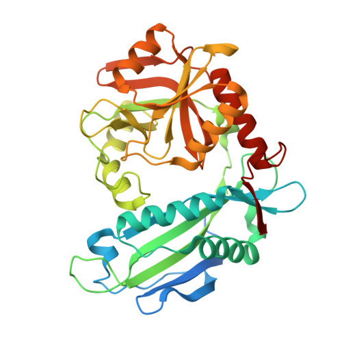
    Unlike mammals, bacteria encode enzymes that synthesize branched-chain amino acids. The pyridoxal 50-phosphate-dependent transaminase performs the final biosynthetic step in these pathways, converting keto acid precursors into -amino acids. The branched-chain amino-acid transaminase from Mycobacterium tuberculosis (MtIlvE) has been crystallized and its structure has been solved at 1.9 angstrom resolution. The MtIlvE monomer is composed of two domains that interact to form the active site. The biologically active form of IlvE is a homodimer in which each monomer contributes a substrate-specificity loop to the partner molecule. Additional substrate selectivity may be imparted by a conserved N-terminal Phe30 residue, which has previously been observed to shield the active site in the type IV fold homodimer. The active site of MtIlvE contains density corresponding to bound PMP, which is likely to be a consequence of the presence of tryptone in the crystallization medium. Additionally, two cysteine residues are positioned at the dimer interface for disulfide-bond formation under oxidative conditions. It is unknown whether they are involved in any regulatory activities analogous to those of the human mitochondrial branched-chain amino-acid transaminase.


  • Organizational Affiliation

    Department of Biochemistry, Albert Einstein College of Medicine, Bronx, NY 10461, USA.


Macromolecules
Find similar proteins by:  (by identity cutoff)  |  3D Structure
Entity ID: 1
MoleculeChains Sequence LengthOrganismDetailsImage
branched-chain-amino-acid aminotransferase368Mycobacterium tuberculosisMutation(s): 0 
Gene Names: ilvEMT2266MTCY190.21cRv2210c
EC: 2.6.1.42
UniProt
Find proteins for P9WQ75 (Mycobacterium tuberculosis (strain ATCC 25618 / H37Rv))
Explore P9WQ75 
Go to UniProtKB:  P9WQ75
Entity Groups  
Sequence Clusters30% Identity50% Identity70% Identity90% Identity95% Identity100% Identity
UniProt GroupP9WQ75
Sequence Annotations
Expand
  • Reference Sequence
Small Molecules
Ligands 1 Unique
IDChains Name / Formula / InChI Key2D Diagram3D Interactions
PMP
Query on PMP

Download Ideal Coordinates CCD File 
B [auth A]4'-DEOXY-4'-AMINOPYRIDOXAL-5'-PHOSPHATE
C8 H13 N2 O5 P
ZMJGSOSNSPKHNH-UHFFFAOYSA-N
Experimental Data & Validation

Experimental Data

  • Method: X-RAY DIFFRACTION
  • Resolution: 1.90 Å
  • R-Value Free: 0.198 
  • R-Value Work: 0.184 
  • R-Value Observed: 0.185 
  • Space Group: P 21 21 2
Unit Cell:
Length ( Å )Angle ( ˚ )
a = 65.454α = 90
b = 85.351β = 90
c = 59.715γ = 90
Software Package:
Software NamePurpose
MOSFLMdata reduction
DENZOdata reduction
SCALEPACKdata scaling
MOLREPphasing
REFMACrefinement
PDB_EXTRACTdata extraction
PHENIXphasing

Structure Validation

View Full Validation Report


Ligand Structure Quality Assessment 


Entry History 

Deposition Data

Revision History  (Full details and data files)

  • Version 1.0: 2009-12-01
    Type: Initial release
  • Version 1.1: 2011-07-13
    Changes: Version format compliance
  • Version 1.2: 2017-11-01
    Changes: Refinement description
  • Version 1.3: 2023-09-06
    Changes: Data collection, Database references, Derived calculations, Refinement description