3HSB

Crystal structure of YmaH (Hfq) from Bacillus subtilis in complex with an RNA aptamer


Experimental Data Snapshot

  • Method: X-RAY DIFFRACTION
  • Resolution: 2.20 Å
  • R-Value Free: 0.228 
  • R-Value Work: 0.214 
  • R-Value Observed: 0.214 

wwPDB Validation   3D Report Full Report


This is version 1.3 of the entry. See complete history


Literature

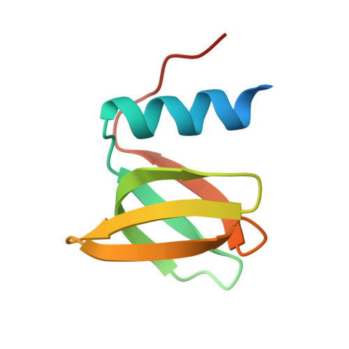
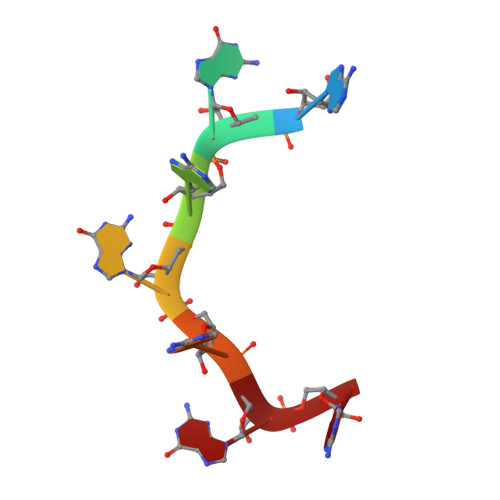
Crystal structure of Hfq from Bacillus subtilis in complex with SELEX-derived RNA aptamer: insight into RNA-binding properties of bacterial Hfq

Someya, T.Baba, S.Fujimoto, M.Kawai, G.Kumasaka, T.Nakamura, K.

(2012) Nucleic Acids Res 40: 1856-1867

  • DOI: https://doi.org/10.1093/nar/gkr892
  • Primary Citation of Related Structures:  
    3HSB

  • PubMed Abstract: 

    Bacterial Hfq is a protein that plays an important role in the regulation of genes in cooperation with sRNAs. Escherichia coli Hfq (EcHfq) has two or more sites that bind RNA(s) including U-rich and/or the poly(A) tail of mRNA. However, functional and structural information about Bacillus subtilis Hfq (BsHfq) including the RNA sequences that specifically bind to it remain unknown. Here, we describe RNA aptamers including fragment (AG)(3)A that are recognized by BsHfq and crystal structures of the BsHfq-(AG)(3)A complex at 2.2 Å resolution. Mutational and structural studies revealed that the RNA fragment binds to the distal site, one of the two binding sites on Hfq, and identified amino acid residues that are critical for sequence-specific interactions between BsHfq and (AG)(3)A. In particular, R32 appears to interact with G bases in (AG)(3)A. Poly(A) also binds to the distal site of EcHfq, but the overall RNA structure and protein-RNA interaction patterns engaged in the R32 residues of BsHfq-(AG)(3)A differ from those of EcHfq-poly(A). These findings provide novel insight into how the Hfq homologue recognizes RNA.


  • Organizational Affiliation

    Graduate School of Life and Environmental Sciences, University of Tsukuba, 1-1-1 Tennodai, Tsukuba-shi, Ibaraki 305-8572, Japan. tsomeya@biol.tsukuba.ac.jp


Macromolecules

Find similar proteins by:  (by identity cutoff)  |  3D Structure
Entity ID: 1
MoleculeChains Sequence LengthOrganismDetailsImage
Protein hfq
A, B, C, D, E
A, B, C, D, E, F
78Bacillus subtilisMutation(s): 0 
Gene Names: ymaH
UniProt
Find proteins for O31796 (Bacillus subtilis (strain 168))
Explore O31796 
Go to UniProtKB:  O31796
Entity Groups  
Sequence Clusters30% Identity50% Identity70% Identity90% Identity95% Identity100% Identity
UniProt GroupO31796
Sequence Annotations
Expand
  • Reference Sequence

Find similar nucleic acids by:  Sequence   |   3D Structure  

Entity ID: 2
MoleculeChains LengthOrganismImage
RNA (5'-R(*AP*GP*AP*GP*AP*GP*A)-3')G [auth X]7N/A
Sequence Annotations
Expand
  • Reference Sequence
Experimental Data & Validation

Experimental Data

  • Method: X-RAY DIFFRACTION
  • Resolution: 2.20 Å
  • R-Value Free: 0.228 
  • R-Value Work: 0.214 
  • R-Value Observed: 0.214 
  • Space Group: I 4 2 2
Unit Cell:
Length ( Å )Angle ( ˚ )
a = 123.7α = 90
b = 123.7β = 90
c = 119.131γ = 90
Software Package:
Software NamePurpose
BSSdata collection
MOLREPphasing
CNSrefinement
HKL-2000data reduction
HKL-2000data scaling

Structure Validation

View Full Validation Report


Entry History 

Deposition Data

Revision History  (Full details and data files)

  • Version 1.0: 2010-06-23
    Type: Initial release
  • Version 1.1: 2011-07-13
    Changes: Version format compliance
  • Version 1.2: 2012-10-03
    Changes: Database references
  • Version 1.3: 2023-11-01
    Changes: Data collection, Database references, Refinement description