3HLN

Crystal structure of ClpP A153C mutant with inter-heptamer disulfide bonds


Experimental Data Snapshot

  • Method: X-RAY DIFFRACTION
  • Resolution: 3.20 Å
  • R-Value Free: 0.253 
  • R-Value Work: 0.213 
  • R-Value Observed: 0.215 

wwPDB Validation   3D Report Full Report


This is version 1.3 of the entry. See complete history


Literature

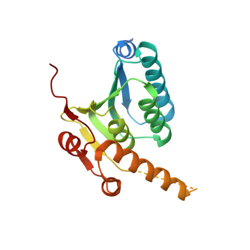
Structural and Theoretical Studies Indicate that the Cylindrical Protease ClpP Samples Extended and Compact Conformations.

Kimber, M.S.Yu, A.Y.Borg, M.Leung, E.Chan, H.S.Houry, W.A.

(2010) Structure 18: 798-808

  • DOI: https://doi.org/10.1016/j.str.2010.04.008
  • Primary Citation of Related Structures:  
    3HLN

  • PubMed Abstract: 

    The highly conserved ClpP protease consists of two heptameric rings that interact by the interdigitation of an alpha-helix beta strand handle domain motif to form a tetradecameric cylinder. We previously proposed that protease dynamics results in the temporary unstructuring of interacting pairs of handle domains, opening transient equatorial side pores that allow for peptide egress. Here, we report the structure of an Escherichia coli ClpP mutant in which each opposing pair of protomers is linked by a disulfide bond. This structure resembles the compact structures of Streptococcus pneumoniae, Mycobacterium tuberculosis, and Plasmodium falciparum ClpPs, rather than the active, extended structures that have previously been determined for E. coli ClpPs. The structural data, along with normal mode analysis, support a model whereby the ClpP cylinder switches dynamically between an active extended state required for substrate degradation and an inactive compact state allowing peptide product release.


  • Organizational Affiliation

    Department of Molecular and Cellular Biology, University of Guelph, Guelph, ON N1G 2W1, Canada.


Macromolecules
Find similar proteins by:  (by identity cutoff)  |  3D Structure
Entity ID: 1
MoleculeChains Sequence LengthOrganismDetailsImage
ATP-dependent Clp protease proteolytic subunit
A,
AA [auth 1],
B,
BA [auth 2],
C,
D,
E,
F,
G,
H,
I,
J,
K,
L,
M,
N,
O,
P,
Q,
R,
S,
T,
U,
V,
W,
X,
Y,
Z
193Escherichia coli K-12Mutation(s): 1 
Gene Names: b0437clpPJW0427lopP
EC: 3.4.21.92
UniProt
Find proteins for P0A6G7 (Escherichia coli (strain K12))
Explore P0A6G7 
Go to UniProtKB:  P0A6G7
Entity Groups  
Sequence Clusters30% Identity50% Identity70% Identity90% Identity95% Identity100% Identity
UniProt GroupP0A6G7
Sequence Annotations
Expand
  • Reference Sequence
Small Molecules
Ligands 1 Unique
IDChains Name / Formula / InChI Key2D Diagram3D Interactions
CA
Query on CA

Download Ideal Coordinates CCD File 
CA [auth E]
DA [auth E]
EA [auth I]
FA [auth I]
GA [auth J]
CA [auth E],
DA [auth E],
EA [auth I],
FA [auth I],
GA [auth J],
HA [auth J],
IA [auth J],
JA [auth M],
KA [auth T],
LA [auth V],
MA [auth Z],
NA [auth 1],
OA [auth 2]
CALCIUM ION
Ca
BHPQYMZQTOCNFJ-UHFFFAOYSA-N
Experimental Data & Validation

Experimental Data

  • Method: X-RAY DIFFRACTION
  • Resolution: 3.20 Å
  • R-Value Free: 0.253 
  • R-Value Work: 0.213 
  • R-Value Observed: 0.215 
  • Space Group: P 32 2 1
Unit Cell:
Length ( Å )Angle ( ˚ )
a = 182.299α = 90
b = 182.299β = 90
c = 476.859γ = 120
Software Package:
Software NamePurpose
ADSCdata collection
MOLREPphasing
REFMACrefinement
HKL-3000data reduction
HKL-3000data scaling

Structure Validation

View Full Validation Report


Entry History 

Deposition Data

Revision History  (Full details and data files)

  • Version 1.0: 2010-07-28
    Type: Initial release
  • Version 1.1: 2011-07-13
    Changes: Source and taxonomy, Version format compliance
  • Version 1.2: 2021-10-13
    Changes: Database references, Derived calculations
  • Version 1.3: 2023-09-06
    Changes: Data collection, Refinement description