3HLM

Crystal Structure of Mouse Mitochondrial Aspartate Aminotransferase/Kynurenine Aminotransferase IV


Experimental Data Snapshot

  • Method: X-RAY DIFFRACTION
  • Resolution: 2.50 Å
  • R-Value Free: 0.234 
  • R-Value Work: 0.181 
  • R-Value Observed: 0.184 

wwPDB Validation   3D Report Full Report


This is version 1.3 of the entry. See complete history


Literature

Structure, expression, and function of kynurenine aminotransferases in human and rodent brains.

Han, Q.Cai, T.Tagle, D.A.Li, J.

(2010) Cell Mol Life Sci 67: 353-368

  • DOI: https://doi.org/10.1007/s00018-009-0166-4
  • Primary Citation of Related Structures:  
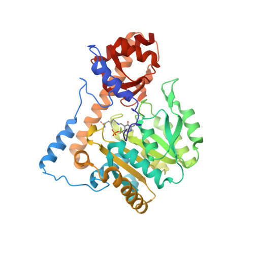
    3HLM

  • PubMed Abstract: 

    Kynurenine aminotransferases (KATs) catalyze the synthesis of kynurenic acid (KYNA), an endogenous antagonist of N-methyl-D: -aspartate and alpha 7-nicotinic acetylcholine receptors. Abnormal KYNA levels in human brains are implicated in the pathophysiology of schizophrenia, Alzheimer's disease, and other neurological disorders. Four KATs have been reported in mammalian brains, KAT I/glutamine transaminase K/cysteine conjugate beta-lyase 1, KAT II/aminoadipate aminotransferase, KAT III/cysteine conjugate beta-lyase 2, and KAT IV/glutamic-oxaloacetic transaminase 2/mitochondrial aspartate aminotransferase. KAT II has a striking tertiary structure in N-terminal part and forms a new subgroup in fold type I aminotransferases, which has been classified as subgroup Iepsilon. Knowledge regarding KATs is vast and complex; therefore, this review is focused on recent important progress of their gene characterization, physiological and biochemical function, and structural properties. The biochemical differences of four KATs, specific enzyme activity assays, and the structural insights into the mechanism of catalysis and inhibition of these enzymes are discussed.


  • Organizational Affiliation

    Department of Biochemistry, Virginia Tech, Blacksburg, VA 24061, USA.


Macromolecules
Find similar proteins by:  (by identity cutoff)  |  3D Structure
Entity ID: 1
MoleculeChains Sequence LengthOrganismDetailsImage
Aspartate aminotransferase, mitochondrial
A, B, C, D
401Mus musculusMutation(s): 0 
Gene Names: Got-2Got2
EC: 2.6.1.1
UniProt & NIH Common Fund Data Resources
Find proteins for P05202 (Mus musculus)
Explore P05202 
Go to UniProtKB:  P05202
IMPC:  MGI:95792
Entity Groups  
Sequence Clusters30% Identity50% Identity70% Identity90% Identity95% Identity100% Identity
UniProt GroupP05202
Sequence Annotations
Expand
  • Reference Sequence
Experimental Data & Validation

Experimental Data

  • Method: X-RAY DIFFRACTION
  • Resolution: 2.50 Å
  • R-Value Free: 0.234 
  • R-Value Work: 0.181 
  • R-Value Observed: 0.184 
  • Space Group: P 21 21 2
Unit Cell:
Length ( Å )Angle ( ˚ )
a = 284.322α = 90
b = 76.792β = 90
c = 87.416γ = 90
Software Package:
Software NamePurpose
HKL-2000data collection
PHASESphasing
REFMACrefinement
HKL-2000data reduction
HKL-2000data scaling

Structure Validation

View Full Validation Report


Entry History 

Deposition Data

Revision History  (Full details and data files)

  • Version 1.0: 2010-06-02
    Type: Initial release
  • Version 1.1: 2011-07-13
    Changes: Version format compliance
  • Version 1.2: 2023-09-06
    Changes: Data collection, Database references, Derived calculations, Refinement description
  • Version 1.3: 2023-11-22
    Changes: Data collection