3HJH

A rigid N-terminal clamp restrains the motor domains of the bacterial transcription-repair coupling factor


Experimental Data Snapshot

  • Method: X-RAY DIFFRACTION
  • Resolution: 1.95 Å
  • R-Value Free: 0.257 
  • R-Value Work: 0.210 
  • R-Value Observed: 0.213 

wwPDB Validation   3D Report Full Report


This is version 1.2 of the entry. See complete history


Literature

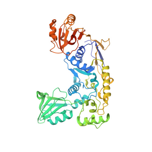
An N-terminal clamp restrains the motor domains of the bacterial transcription-repair coupling factor Mfd.

Murphy, M.N.Gong, P.Ralto, K.Manelyte, L.Savery, N.J.Theis, K.

(2009) Nucleic Acids Res 37: 6042-6053

  • DOI: https://doi.org/10.1093/nar/gkp680
  • Primary Citation of Related Structures:  
    3HJH

  • PubMed Abstract: 

    Motor proteins that translocate on nucleic acids are key players in gene expression and maintenance. While the function of these proteins is diverse, they are driven by highly conserved core motor domains. In transcription-coupled DNA repair, motor activity serves to remove RNA polymerase stalled on damaged DNA, making the lesion accessible for repair. Structural and biochemical data on the bacterial transcription-repair coupling factor Mfd suggest that this enzyme undergoes large conformational changes from a dormant state to an active state upon substrate binding. Mfd can be functionally dissected into an N-terminal part instrumental in recruiting DNA repair proteins (domains 1-3, MfdN), and a C-terminal part harboring motor activity (domains 4-7, MfdC). We show that isolated MfdC has elevated ATPase and motor activities compared to the full length protein. While MfdN has large effects on MfdC activity and thermostability in cis, these effects are not observed in trans. The structure of MfdN is independent of interactions with MfdC, implying that MfdN acts as a clamp that restrains motions of the motor domains in the dormant state. We conclude that releasing MfdN:MfdC interactions serves as a central molecular switch that upregulates Mfd functions during transcription-coupled DNA repair.


  • Organizational Affiliation

    Department of Chemistry, Department of Biochemistry & Molecular Biology, University of Massachusetts Amherst, Amherst, Massachusetts 01003, USA.


Macromolecules
Find similar proteins by:  (by identity cutoff)  |  3D Structure
Entity ID: 1
MoleculeChains Sequence LengthOrganismDetailsImage
Transcription-repair-coupling factor483Escherichia coli K-12Mutation(s): 0 
Gene Names: b1114JW1100mfd
EC: 3.6.1
UniProt
Find proteins for P30958 (Escherichia coli (strain K12))
Explore P30958 
Go to UniProtKB:  P30958
Entity Groups  
Sequence Clusters30% Identity50% Identity70% Identity90% Identity95% Identity100% Identity
UniProt GroupP30958
Sequence Annotations
Expand
  • Reference Sequence
Small Molecules
Ligands 1 Unique
IDChains Name / Formula / InChI Key2D Diagram3D Interactions
CO
Query on CO

Download Ideal Coordinates CCD File 
B [auth A]COBALT (II) ION
Co
XLJKHNWPARRRJB-UHFFFAOYSA-N
Experimental Data & Validation

Experimental Data

  • Method: X-RAY DIFFRACTION
  • Resolution: 1.95 Å
  • R-Value Free: 0.257 
  • R-Value Work: 0.210 
  • R-Value Observed: 0.213 
  • Space Group: P 21 21 2
Unit Cell:
Length ( Å )Angle ( ˚ )
a = 84.062α = 90
b = 157.65β = 90
c = 35.964γ = 90
Software Package:
Software NamePurpose
ADSCdata collection
MOLREPphasing
REFMACrefinement
HKL-2000data reduction
HKL-2000data scaling

Structure Validation

View Full Validation Report


Entry History 

Deposition Data

Revision History  (Full details and data files)

  • Version 1.0: 2009-10-20
    Type: Initial release
  • Version 1.1: 2011-07-13
    Changes: Advisory, Version format compliance
  • Version 1.2: 2023-09-06
    Changes: Data collection, Database references, Derived calculations, Refinement description