3HBB

Structures of dihydrofolate reductase-thymidylate synthase of Trypanosoma cruzi in the folate-free state and in complex with two antifolate drugs, trimetrexate and methotrexate


Experimental Data Snapshot

  • Method: X-RAY DIFFRACTION
  • Resolution: 3.00 Å
  • R-Value Free: 0.239 
  • R-Value Work: 0.201 
  • R-Value Observed: 0.203 

wwPDB Validation   3D Report Full Report


Ligand Structure Quality Assessment 


This is version 1.2 of the entry. See complete history


Literature

Structures of dihydrofolate reductase-thymidylate synthase of Trypanosoma cruzi in the folate-free state and in complex with two antifolate drugs, trimetrexate and methotrexate.

Senkovich, O.Schormann, N.Chattopadhyay, D.

(2009) Acta Crystallogr D Biol Crystallogr 65: 704-716

  • DOI: https://doi.org/10.1107/S090744490901230X
  • Primary Citation of Related Structures:  
    3HBB

  • PubMed Abstract: 

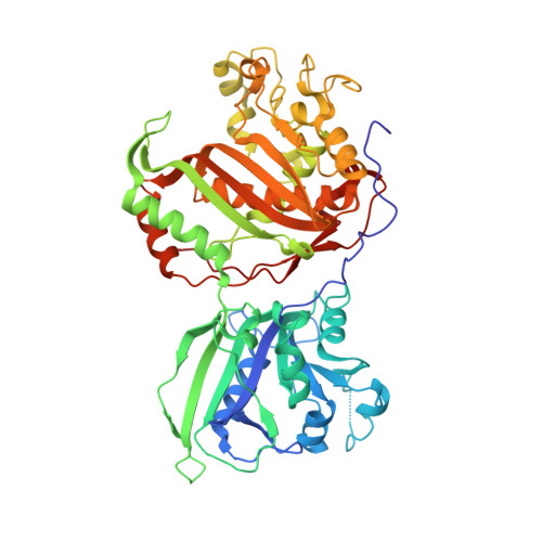
    The flagellate protozoan parasite Trypanosoma cruzi is the pathogenic agent of Chagas disease (also called American trypanosomiasis), which causes approximately 50,000 deaths annually. The disease is endemic in South and Central America. The parasite is usually transmitted by a blood-feeding insect vector, but can also be transmitted via blood transfusion. In the chronic form, Chagas disease causes severe damage to the heart and other organs. There is no satisfactory treatment for chronic Chagas disease and no vaccine is available. There is an urgent need for the development of chemotherapeutic agents for the treatment of T. cruzi infection and therefore for the identification of potential drug targets. The dihydrofolate reductase activity of T. cruzi, which is expressed as part of a bifunctional enzyme, dihydrofolate reductase-thymidylate synthase (DHFR-TS), is a potential target for drug development. In order to gain a detailed understanding of the structure-function relationship of T. cruzi DHFR, the three-dimensional structure of this protein in complex with various ligands is being studied. Here, the crystal structures of T. cruzi DHFR-TS with three different compositions of the DHFR domain are reported: the folate-free state, the complex with the lipophilic antifolate trimetrexate (TMQ) and the complex with the classical antifolate methotrexate (MTX). These structures reveal that the enzyme is a homodimer with substantial interactions between the two TS domains of neighboring subunits. In contrast to the enzymes from Cryptosporidium hominis and Plasmodium falciparum, the DHFR and TS active sites of T. cruzi lie on the same side of the monomer. As in other parasitic DHFR-TS proteins, the N-terminal extension of the T. cruzi enzyme is involved in extensive interactions between the two domains. The DHFR active site of the T. cruzi enzyme shows subtle differences compared with its human counterpart. These differences may be exploited for the development of antifolate-based therapeutic agents for the treatment of T. cruzi infection.


  • Organizational Affiliation

    Center for Biophysical Sciences and Engineering, University of Alabama at Birmingham, Birmingham, AL 35294, USA.


Macromolecules
Find similar proteins by:  (by identity cutoff)  |  3D Structure
Entity ID: 1
MoleculeChains Sequence LengthOrganismDetailsImage
Dihydrofolate reductase-thymidylate synthase
A, B, C, D
521Trypanosoma cruziMutation(s): 0 
Gene Names: dhfr-ts
EC: 1.5.1.3 (PDB Primary Data), 2.1.1.45 (PDB Primary Data)
UniProt
Find proteins for Q27793 (Trypanosoma cruzi)
Explore Q27793 
Go to UniProtKB:  Q27793
Entity Groups  
Sequence Clusters30% Identity50% Identity70% Identity90% Identity95% Identity100% Identity
UniProt GroupQ27793
Sequence Annotations
Expand
  • Reference Sequence
Small Molecules
Ligands 4 Unique
IDChains Name / Formula / InChI Key2D Diagram3D Interactions
NAP
Query on NAP

Download Ideal Coordinates CCD File 
E [auth A],
LA [auth D],
O [auth B],
X [auth C]
NADP NICOTINAMIDE-ADENINE-DINUCLEOTIDE PHOSPHATE
C21 H28 N7 O17 P3
XJLXINKUBYWONI-NNYOXOHSSA-N
TMQ
Query on TMQ

Download Ideal Coordinates CCD File 
F [auth A],
MA [auth D],
P [auth B],
Y [auth C]
TRIMETREXATE
C19 H24 N5 O3
NOYPYLRCIDNJJB-UHFFFAOYSA-O
SO4
Query on SO4

Download Ideal Coordinates CCD File 
AA [auth C]
BA [auth C]
CA [auth C]
DA [auth C]
EA [auth C]
AA [auth C],
BA [auth C],
CA [auth C],
DA [auth C],
EA [auth C],
FA [auth C],
G [auth A],
GA [auth C],
H [auth A],
I [auth A],
J [auth A],
K [auth A],
L [auth A],
NA [auth D],
OA [auth D],
PA [auth D],
Q [auth B],
QA [auth D],
R [auth B],
RA [auth D],
S [auth B],
SA [auth D],
T [auth B],
TA [auth D],
U [auth B],
UA [auth D],
W [auth C],
Z [auth C]
SULFATE ION
O4 S
QAOWNCQODCNURD-UHFFFAOYSA-L
EDO
Query on EDO

Download Ideal Coordinates CCD File 
HA [auth C]
IA [auth C]
JA [auth C]
KA [auth C]
M [auth A]
HA [auth C],
IA [auth C],
JA [auth C],
KA [auth C],
M [auth A],
N [auth A],
V [auth B],
VA [auth D],
WA [auth D],
XA [auth D],
YA [auth D],
ZA [auth D]
1,2-ETHANEDIOL
C2 H6 O2
LYCAIKOWRPUZTN-UHFFFAOYSA-N
Experimental Data & Validation

Experimental Data

  • Method: X-RAY DIFFRACTION
  • Resolution: 3.00 Å
  • R-Value Free: 0.239 
  • R-Value Work: 0.201 
  • R-Value Observed: 0.203 
  • Space Group: P 43 21 2
Unit Cell:
Length ( Å )Angle ( ˚ )
a = 176.571α = 90
b = 176.571β = 90
c = 251.848γ = 90
Software Package:
Software NamePurpose
HKL-2000data collection
PHASERphasing
REFMACrefinement
d*TREKdata reduction
d*TREKdata scaling

Structure Validation

View Full Validation Report


Ligand Structure Quality Assessment 


Entry History 

Deposition Data

Revision History  (Full details and data files)

  • Version 1.0: 2009-05-19
    Type: Initial release
  • Version 1.1: 2011-07-13
    Changes: Advisory, Version format compliance
  • Version 1.2: 2023-09-06
    Changes: Data collection, Database references, Derived calculations, Refinement description