3H5R

Crystal structure of E. coli MccB + Succinimide


Experimental Data Snapshot

  • Method: X-RAY DIFFRACTION
  • Resolution: 2.10 Å
  • R-Value Free: 0.250 
  • R-Value Work: 0.194 
  • R-Value Observed: 0.197 

wwPDB Validation   3D Report Full Report


This is version 2.0 of the entry. See complete history


Literature

How the MccB bacterial ancestor of ubiquitin E1 initiates biosynthesis of the microcin C7 antibiotic.

Regni, C.A.Roush, R.F.Miller, D.J.Nourse, A.Walsh, C.T.Schulman, B.A.

(2009) EMBO J 28: 1953-1964

  • DOI: https://doi.org/10.1038/emboj.2009.146
  • Primary Citation of Related Structures:  
    3H5A, 3H5N, 3H5R, 3H9G, 3H9J, 3H9Q

  • PubMed Abstract: 

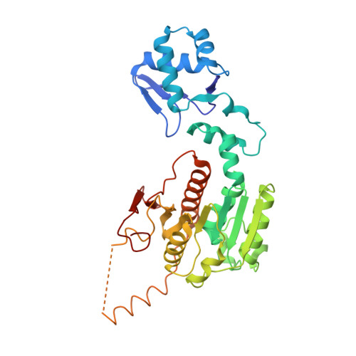
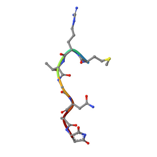
    The 39-kDa Escherichia coli enzyme MccB catalyses a remarkable posttranslational modification of the MccA heptapeptide during the biosynthesis of microcin C7 (MccC7), a 'Trojan horse' antibiotic. The approximately 260-residue C-terminal region of MccB is homologous to ubiquitin-like protein (UBL) activating enzyme (E1) adenylation domains. Accordingly, MccB-catalysed C-terminal MccA-acyl-adenylation is reminiscent of the E1-catalysed activation reaction. However, unlike E1 substrates, which are UBLs with a C-terminal di-glycine sequence, MccB's substrate, MccA, is a short peptide with an essential C-terminal Asn. Furthermore, after an intramolecular rearrangement of MccA-acyl-adenylate, MccB catalyses a second, unique reaction, producing a stable phosphoramidate-linked analogue of acyl-adenylated aspartic acid. We report six-crystal structures of MccB in apo, substrate-, intermediate-, and inhibitor-bound forms. Structural and kinetic analyses reveal a novel-peptide clamping mechanism for MccB binding to heptapeptide substrates and a dynamic-active site for catalysing dual adenosine triphosphate-consuming reactions. The results provide insight into how a distinctive member of the E1 superfamily carries out two-step activation for generating the peptidyl-antibiotic MccC7.


  • Organizational Affiliation

    Department of Structural Biology, St Jude Children's Research Hospital, Memphis, TN 38105, USA.


Macromolecules
Find similar proteins by:  (by identity cutoff)  |  3D Structure
Entity ID: 1
MoleculeChains Sequence LengthOrganismDetailsImage
MccB protein
A, B, C, D
353Escherichia coliMutation(s): 0 
Gene Names: mccB
UniProt
Find proteins for Q47506 (Escherichia coli)
Explore Q47506 
Go to UniProtKB:  Q47506
Entity Groups  
Sequence Clusters30% Identity50% Identity70% Identity90% Identity95% Identity100% Identity
UniProt GroupQ47506
Sequence Annotations
Expand
  • Reference Sequence

Find similar proteins by:  Sequence   |   3D Structure  

Entity ID: 2
MoleculeChains Sequence LengthOrganismDetailsImage
Microcin C7 analog
E, F, G, H
7Escherichia coliMutation(s): 0 
UniProt
Find proteins for Q47505 (Escherichia coli)
Explore Q47505 
Go to UniProtKB:  Q47505
Entity Groups  
UniProt GroupQ47505
Sequence Annotations
Expand
  • Reference Sequence
Biologically Interesting Molecules (External Reference) 1 Unique
Experimental Data & Validation

Experimental Data

  • Method: X-RAY DIFFRACTION
  • Resolution: 2.10 Å
  • R-Value Free: 0.250 
  • R-Value Work: 0.194 
  • R-Value Observed: 0.197 
  • Space Group: P 1 21 1
Unit Cell:
Length ( Å )Angle ( ˚ )
a = 55.934α = 90
b = 137.972β = 92.1
c = 80.134γ = 90
Software Package:
Software NamePurpose
DENZOdata reduction
SCALEPACKdata scaling
REFMACrefinement
PDB_EXTRACTdata extraction
X-GENdata scaling
HKL-2000data reduction
REFMACphasing

Structure Validation

View Full Validation Report


Entry History 

Deposition Data

Revision History  (Full details and data files)

  • Version 1.0: 2009-06-16
    Type: Initial release
  • Version 1.1: 2011-07-13
    Changes: Version format compliance
  • Version 1.2: 2011-07-27
    Changes: Non-polymer description, Structure summary
  • Version 1.3: 2012-12-12
    Changes: Other
  • Version 1.4: 2017-11-01
    Changes: Refinement description
  • Version 1.5: 2023-09-06
    Changes: Data collection, Database references, Derived calculations, Refinement description
  • Version 2.0: 2023-11-15
    Changes: Atomic model, Data collection, Derived calculations