Structural basis for resistance of the genotype 2b hepatitis C virus NS5B polymerase to site A non-nucleoside inhibitors.
Rydberg, E.H., Cellucci, A., Bartholomew, L., Mattu, M., Barbato, G., Ludmerer, S.W., Graham, D.J., Altamura, S., Paonessa, G., De Francesco, R., Migliaccio, G., Carfi, A.(2009) J Mol Biol 390: 1048-1059
- PubMed: 19505479
- DOI: https://doi.org/10.1016/j.jmb.2009.06.012
- Primary Citation of Related Structures:
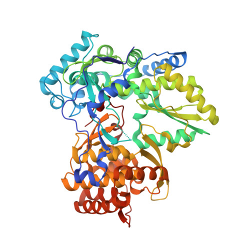
3GSZ - PubMed Abstract:
Hepatitis C virus (HCV) exists in six major genotypes. Compared with the 1b enzyme, genotype 2b HCV polymerase exhibits a more than 100-fold reduction in sensitivity to the indole-N-acetamide class of non-nucleoside inhibitors. These compounds have been shown to bind in a pocket occupied by helix A of the mobile Lambda1 loop in the apoenzyme. The three-dimensional structure of the HCV polymerase from genotype 2b was determined to 1.9-A resolution and compared with the genotype 1b enzyme. This structural analysis suggests that genotypic variants result in a different shape of the inhibitor binding site. Mutants of the inhibitor binding pocket were generated in a 1b enzyme and evaluated for their binding affinity and sensitivity to inhibition by indole-N-acetamides. Most of the point mutants showed little variation in activity and IC(50), with the exception of 15- and 7-fold increases in IC(50) for Leu392Ile and Val494Ala mutants (1b-->2b), respectively. Furthermore, a 1b replicon with 20-fold resistance to this class of inhibitors was selected and shown to contain the Leu392Ile mutation. Chimeric enzymes, where the 2b fingertip Lambda1 loop, pocket or both replaced the corresponding regions of the 1b enzyme, were also generated. The fingertip chimera retained 1b-like inhibitor binding affinity, whereas the other two chimeric constructs and the 2b enzyme displayed between 50- and 100-fold reduction in binding affinity. Together, these data suggest that differences in the amino acid composition and shape of the indole-N-acetamide binding pocket are responsible for the resistance of the 2b polymerase to this class of inhibitors.
Organizational Affiliation:
Istituto di Ricerca di Biologia Molecolare, Rome, Italy.