


Crystal structure of short chain dehydrogenase reductase SDR glucose-ribitol dehydrogenase from Brucella melitensis
Edwards, T.E., Staker, B.L., Seattle Structural Genomics Center for Infectious Disease (SSGCID)To be published.
Experimental Data Snapshot
wwPDB Validation 3D Report Full Report

Entity ID: 1 | |||||
|---|---|---|---|---|---|
| Molecule | Chains | Sequence Length | Organism | Details | Image |
| Enoyl-(acyl-carrier-protein) reductase (NADH) | 293 | Brucella melitensis | Mutation(s): 0 Gene Names: BMEI1512 EC: 1.3.1.9 | 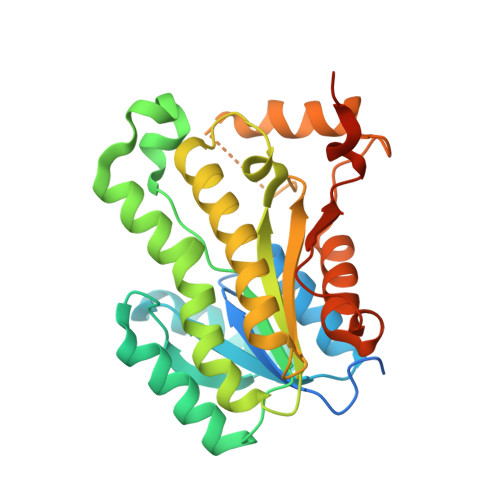 | |
UniProt | |||||
Find proteins for Q8YFK8 (Brucella melitensis biotype 1 (strain 16M / ATCC 23456 / NCTC 10094)) Explore Q8YFK8 Go to UniProtKB: Q8YFK8 | |||||
Entity Groups | |||||
| Sequence Clusters | 30% Identity50% Identity70% Identity90% Identity95% Identity100% Identity | ||||
| UniProt Group | Q8YFK8 | ||||
Sequence AnnotationsExpand | |||||
| |||||
| Length ( Å ) | Angle ( ˚ ) |
|---|---|
| a = 66.1 | α = 90 |
| b = 120.1 | β = 99.2 |
| c = 137.7 | γ = 90 |
| Software Name | Purpose |
|---|---|
| XSCALE | data scaling |
| MOLREP | phasing |
| REFMAC | refinement |
| PDB_EXTRACT | data extraction |
RCSB PDB (citation) is hosted by
RCSB PDB is a member of the

