Crystal structure of the NADP-dependent mannitol dehydrogenase from Cladosporium herbarum: Implications for oligomerisation and catalysis.
Nuss, D., Goettig, P., Magler, I., Denk, U., Breitenbach, M., Schneider, P.B., Brandstetter, H., Simon-Nobbe, B.(2010) Biochimie 92: 985-993
- PubMed: 20420880
- DOI: https://doi.org/10.1016/j.biochi.2010.04.012
- Primary Citation of Related Structures:
3GDF, 3GDG - PubMed Abstract:
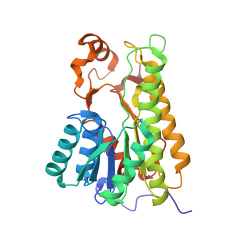
The ascomycete Cladosporium herbarum is a prominent fungal inducer of Type I allergy. The only major allergen identified so far is Cla h 8, a NADP-dependent mannitol dehydrogenase (MtDH). MtDH, a cytoplasmic protein of 28.5kDa, belongs to the Short chain Dehydrogenases/Reductases (SDR), acting as a NADP-dependent oxidoreductase. In this study, we found that C. herbarum MtDH can exist as monomers, dimers and tetramers in solution and, correspondingly, forms tetramers and higher oligomers in two crystal structures. Additionally, we identified a unique adaptive binding site for the metal ions Na(+) and Zn(2+) that were distinguished by an anomalous dispersion experiment. A Translation-Libration-Screw analysis confirmed the stabilising effect of Zn(2+) for the tetrameric assembly. Moreover, the zinc containing structure explains the mode of MtDH multimerisation by metal bridging of the tetramers. The formation of oligomers and higher multimers of MtDH provides a missing link to its allergenic properties. Based on the well defined active site region and a comparative analysis with related structures, we can also clarify the atypical enzymatic properties of MtDH by two alternative binding modes of the substrate to the active site.
Organizational Affiliation:
Division of Structural Biology, Department of Molecular Biology, University of Salzburg, Salzburg, Austria. Dorota.Nuess@sbg.ac.at