3G5I

Crystal Structure of the E.coli RihA pyrimidine nucleosidase bound to a iminoribitol-based inhibitor


Experimental Data Snapshot

  • Method: X-RAY DIFFRACTION
  • Resolution: 2.10 Å
  • R-Value Free: 0.241 
  • R-Value Work: 0.203 
  • R-Value Observed: 0.205 

wwPDB Validation   3D Report Full Report


Ligand Structure Quality Assessment 


This is version 1.3 of the entry. See complete history


Literature

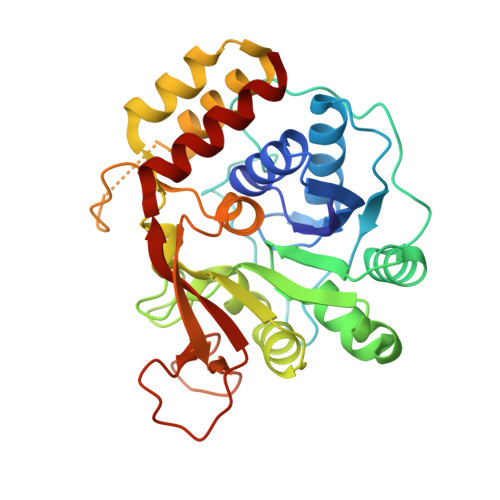
Active site plasticity revealed from the structure of the enterobacterial N-ribohydrolase RihA bound to a competitive inhibitor.

Garau, G.Muzzolini, L.Tornaghi, P.Degano, M.

(2010) BMC Struct Biol 10: 14-14

  • DOI: https://doi.org/10.1186/1472-6807-10-14
  • Primary Citation of Related Structures:  
    3G5I

  • PubMed Abstract: 

    Pyrimidine-preferring N-ribohydrolases (CU-NHs) are a class of Ca2+-dependent enzymes that catalyze the hydrolytic cleavage of the N-glycosidic bond in pyrimidine nucleosides. With the exception of few selected organisms, their physiological relevance in prokaryotes and eukaryotes is yet under investigation.


  • Organizational Affiliation

    Biocrystallography Unit, Division of Immunology, Transplantation, and Infectious Diseases - Scientific Institute S. Raffaele, via Olgettina 58, 20132 Milan - Italy.


Macromolecules
Find similar proteins by:  (by identity cutoff)  |  3D Structure
Entity ID: 1
MoleculeChains Sequence LengthOrganismDetailsImage
Pyrimidine-specific ribonucleoside hydrolase rihA
A, B, C, D
312Escherichia coli K-12Mutation(s): 0 
Gene Names: ybeK
EC: 3.2.2.8
UniProt
Find proteins for P41409 (Escherichia coli (strain K12))
Explore P41409 
Go to UniProtKB:  P41409
Entity Groups  
Sequence Clusters30% Identity50% Identity70% Identity90% Identity95% Identity100% Identity
UniProt GroupP41409
Sequence Annotations
Expand
  • Reference Sequence
Small Molecules
Ligands 3 Unique
IDChains Name / Formula / InChI Key2D Diagram3D Interactions
DNB
Query on DNB

Download Ideal Coordinates CCD File 
G [auth A],
I [auth B],
K [auth C],
M [auth D]
(2S,3S,4R,5R)-2-(3,4-diaminophenyl)-5-(hydroxymethyl)pyrrolidine-3,4-diol
C11 H17 N3 O3
YAYMFJWCRXEXGZ-YTWAJWBKSA-N
BME
Query on BME

Download Ideal Coordinates CCD File 
E [auth A]BETA-MERCAPTOETHANOL
C2 H6 O S
DGVVWUTYPXICAM-UHFFFAOYSA-N
CA
Query on CA

Download Ideal Coordinates CCD File 
F [auth A],
H [auth B],
J [auth C],
L [auth D]
CALCIUM ION
Ca
BHPQYMZQTOCNFJ-UHFFFAOYSA-N
Experimental Data & Validation

Experimental Data

  • Method: X-RAY DIFFRACTION
  • Resolution: 2.10 Å
  • R-Value Free: 0.241 
  • R-Value Work: 0.203 
  • R-Value Observed: 0.205 
  • Space Group: P 1 21 1
Unit Cell:
Length ( Å )Angle ( ˚ )
a = 84.35α = 90
b = 82.91β = 112.15
c = 93.87γ = 90
Software Package:
Software NamePurpose
DNAdata collection
MOLREPphasing
REFMACrefinement
XDSdata reduction
XSCALEdata scaling

Structure Validation

View Full Validation Report


Ligand Structure Quality Assessment 


Entry History 

Deposition Data

Revision History  (Full details and data files)

  • Version 1.0: 2010-02-09
    Type: Initial release
  • Version 1.1: 2011-07-13
    Changes: Advisory, Version format compliance
  • Version 1.2: 2021-02-17
    Changes: Database references, Derived calculations
  • Version 1.3: 2023-11-01
    Changes: Data collection, Database references, Refinement description