3FR8

rice Ketolacid reductoisomerase in complex with Mg2+-NADPH


Experimental Data Snapshot

  • Method: X-RAY DIFFRACTION
  • Resolution: 2.80 Å
  • R-Value Free: 0.284 
  • R-Value Work: 0.233 
  • R-Value Observed: 0.236 

wwPDB Validation   3D Report Full Report


Ligand Structure Quality Assessment 


This is version 1.3 of the entry. See complete history


Literature

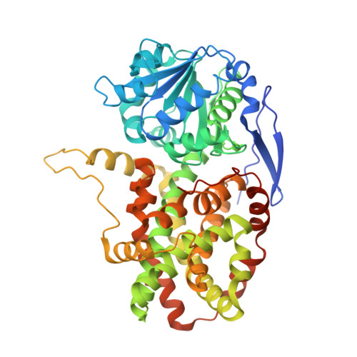
Conformational changes in a plant ketol-acid reductoisomerase upon Mg(2+) and NADPH binding as revealed by two crystal structures

Leung, E.W.Guddat, L.W.

(2009) J Mol Biol 389: 167-182

  • DOI: https://doi.org/10.1016/j.jmb.2009.04.012
  • Primary Citation of Related Structures:  
    3FR7, 3FR8

  • PubMed Abstract: 

    Ketol-acid reductoisomerase (KARI; EC 1.1.1.86) is an enzyme in the branched-chain amino acid biosynthesis pathway where it catalyzes the conversion of 2-acetolactate into (2R)-2,3-dihydroxy-3-isovalerate or the conversion of 2-aceto-2-hydroxybutyrate into (2R,3R)-2,3-dihydroxy-3-methylvalerate. KARI catalyzes two reactions-alkyl migration and reduction-and requires Mg(2+) and NADPH for activity. To date, the only reported structures for a plant KARI are those of the spinach enzyme-Mn(2+)-(phospho)ADP ribose-(2R,3R)-2,3-dihydroxy-3-methylvalerate complex and the spinach KARI-Mg(2)(+)-NADPH-N-hydroxy-N-isopropyloxamate complex, where N-hydroxy-N-isopropyloxamate is a predicted transition-state analog. These studies demonstrated that the enzyme consists of two domains, N-domain and C-domain, with the active site at the interface of these domains. Here, we have determined the structures of the rice KARI-Mg(2+) and rice KARI-Mg(2)(+)-NADPH complexes to 1.55 A and 2.80 A resolutions, respectively. In comparing the structures of all the complexes, several differences are observed. Firstly, the N-domain is rotated up to 15 degrees relative to the C-domain, expanding the active site by up to 4 A. Secondly, an alpha-helix in the C-domain that includes residues V510-T519 and forms part of the active site moves by approximately 3.9 A upon binding of NADPH. Thirdly, the 15 C-terminal amino acid residues in the rice KARI-Mg(2+) complex are disordered. In the rice KARI-Mg(2)(+)-NADPH complex and the spinach KARI structures, many of the 15 residues bind to NADPH and the N-domain and cover the active site. Fourthly, the location of the metal ions within the active site can vary by up to 2.7 A. The new structures allow us to propose that an induced-fit mechanism operates to (i) allow substrate to enter the active site, (ii) close over the active site during catalysis, and (iii) open the active site to facilitate product release.


  • Organizational Affiliation

    The University of Queensland, Brisbane, Australia.


Macromolecules
Find similar proteins by:  (by identity cutoff)  |  3D Structure
Entity ID: 1
MoleculeChains Sequence LengthOrganismDetailsImage
Putative ketol-acid reductoisomerase (Os05g0573700 protein)
A, B
525Oryza sativa Japonica GroupMutation(s): 0 
Gene Names: ilvBOJ1735_C10.18Os05g0573700
EC: 1.1.1.86
UniProt
Find proteins for Q65XK0 (Oryza sativa subsp. japonica)
Explore Q65XK0 
Go to UniProtKB:  Q65XK0
Entity Groups  
Sequence Clusters30% Identity50% Identity70% Identity90% Identity95% Identity100% Identity
UniProt GroupQ65XK0
Sequence Annotations
Expand
  • Reference Sequence
Small Molecules
Ligands 5 Unique
IDChains Name / Formula / InChI Key2D Diagram3D Interactions
NDP
Query on NDP

Download Ideal Coordinates CCD File 
E [auth A],
I [auth B]
NADPH DIHYDRO-NICOTINAMIDE-ADENINE-DINUCLEOTIDE PHOSPHATE
C21 H30 N7 O17 P3
ACFIXJIJDZMPPO-NNYOXOHSSA-N
PE4
Query on PE4

Download Ideal Coordinates CCD File 
F [auth A]2-{2-[2-(2-{2-[2-(2-ETHOXY-ETHOXY)-ETHOXY]-ETHOXY}-ETHOXY)-ETHOXY]-ETHOXY}-ETHANOL
C16 H34 O8
PJWQOENWHPEPKI-UHFFFAOYSA-N
PEG
Query on PEG

Download Ideal Coordinates CCD File 
J [auth B]DI(HYDROXYETHYL)ETHER
C4 H10 O3
MTHSVFCYNBDYFN-UHFFFAOYSA-N
SO4
Query on SO4

Download Ideal Coordinates CCD File 
D [auth A]SULFATE ION
O4 S
QAOWNCQODCNURD-UHFFFAOYSA-L
MG
Query on MG

Download Ideal Coordinates CCD File 
C [auth A],
G [auth B],
H [auth B]
MAGNESIUM ION
Mg
JLVVSXFLKOJNIY-UHFFFAOYSA-N
Experimental Data & Validation

Experimental Data

  • Method: X-RAY DIFFRACTION
  • Resolution: 2.80 Å
  • R-Value Free: 0.284 
  • R-Value Work: 0.233 
  • R-Value Observed: 0.236 
  • Space Group: I 41 2 2
Unit Cell:
Length ( Å )Angle ( ˚ )
a = 162.06α = 90
b = 162.06β = 90
c = 196.47γ = 90
Software Package:
Software NamePurpose
CrystalCleardata collection
AMoREphasing
REFMACrefinement
CrystalCleardata reduction
SCALEPACKdata scaling

Structure Validation

View Full Validation Report


Ligand Structure Quality Assessment 


Entry History 

Deposition Data

Revision History  (Full details and data files)

  • Version 1.0: 2009-04-14
    Type: Initial release
  • Version 1.1: 2011-07-13
    Changes: Advisory, Version format compliance
  • Version 1.2: 2011-11-16
    Changes: Atomic model
  • Version 1.3: 2023-09-06
    Changes: Advisory, Data collection, Database references, Derived calculations, Refinement description