3FO9

Crystal structure of aldolase antibody 33F12 Fab' in complex with hapten 1,3-diketone


Experimental Data Snapshot

  • Method: X-RAY DIFFRACTION
  • Resolution: 1.90 Å
  • R-Value Free: 0.249 
  • R-Value Work: 0.215 
  • R-Value Observed: 0.216 

wwPDB Validation   3D Report Full Report


Ligand Structure Quality Assessment 


This is version 1.3 of the entry. See complete history


Literature

Direct observation of an enamine intermediate in amine catalysis

Zhu, X.Tanaka, F.Lerner, R.A.Barbas, C.F.Wilson, I.A.

(2009) J Am Chem Soc 131: 18206-18207

  • DOI: https://doi.org/10.1021/ja907271a
  • Primary Citation of Related Structures:  
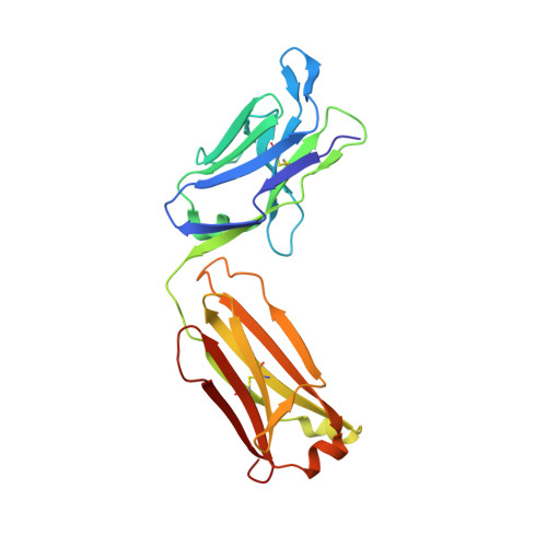
    3FO9

  • PubMed Abstract: 

    An enamine intermediate is believed to be the central feature of biological catalysts, such as aldolases and small molecule amine organocatalysts. Despite decades of investigation of naturally occurring aldolase enzymes and recent studies on designed aldolase antibodies and organocatalysts, direct structural observation of an enamine intermediate has proven to be rare. Herein, we report the observation of a stable enamine intermediate in the crystal structure of an aldolase antibody 33F12 in complex with a 1,3-diketone derivative. This enamine complex structure provides strong evidence that fewer residues are essential for amine catalysis within the hydrophobic environments of this catalytic antibody than speculated for natural aldolase enzymes and should serve to guide future studies aimed at the rational design of these types of catalysts, as well as organocatalysts. Indeed, enamine catalysis in proteins might be more simplistic than previously imagined.


  • Organizational Affiliation

    Department of Chemistry, The Skaggs Institute for Chemical Biology, The Scripps Research Institute, 10550 North Torrey Pines Road, La Jolla, California 92037, USA.


Macromolecules
Find similar proteins by:  (by identity cutoff)  |  3D Structure
Entity ID: 1
MoleculeChains Sequence LengthOrganismDetailsImage
Immunoglobulin IGG2A - light chainA [auth L],
C [auth A]
219Mus musculusMutation(s): 0 
Entity Groups  
Sequence Clusters30% Identity50% Identity70% Identity90% Identity95% Identity100% Identity
Sequence Annotations
Expand
  • Reference Sequence
Find similar proteins by:  (by identity cutoff)  |  3D Structure
Entity ID: 2
MoleculeChains Sequence LengthOrganismDetailsImage
Immunoglobulin IGG2A - heavy chainB [auth H],
D [auth B]
218Mus musculusMutation(s): 0 
UniProt
Find proteins for P01865 (Mus musculus)
Explore P01865 
Go to UniProtKB:  P01865
Entity Groups  
Sequence Clusters30% Identity50% Identity70% Identity90% Identity95% Identity100% Identity
UniProt GroupP01865
Sequence Annotations
Expand
  • Reference Sequence
Small Molecules
Ligands 1 Unique
IDChains Name / Formula / InChI Key2D Diagram3D Interactions
DIK
Query on DIK

Download Ideal Coordinates CCD File 
E [auth H],
F [auth B]
5-{[4-(5-methyl-3-oxohex-4-en-1-yl)phenyl]amino}-5-oxopentanoic acid
C18 H23 N O4
SZAPYZUSRJAHQP-UHFFFAOYSA-N
Experimental Data & Validation

Experimental Data

  • Method: X-RAY DIFFRACTION
  • Resolution: 1.90 Å
  • R-Value Free: 0.249 
  • R-Value Work: 0.215 
  • R-Value Observed: 0.216 
  • Space Group: P 21 21 21
Unit Cell:
Length ( Å )Angle ( ˚ )
a = 65.83α = 90
b = 81.038β = 90
c = 174.314γ = 90
Software Package:
Software NamePurpose
DENZOdata reduction
SCALEPACKdata scaling
AMoREphasing
REFMACrefinement
PDB_EXTRACTdata extraction
HKL-2000data collection

Structure Validation

View Full Validation Report


Ligand Structure Quality Assessment 


Entry History 

Deposition Data

Revision History  (Full details and data files)

  • Version 1.0: 2009-12-22
    Type: Initial release
  • Version 1.1: 2011-07-13
    Changes: Advisory, Version format compliance
  • Version 1.2: 2017-11-01
    Changes: Refinement description
  • Version 1.3: 2023-09-06
    Changes: Data collection, Database references, Derived calculations, Refinement description