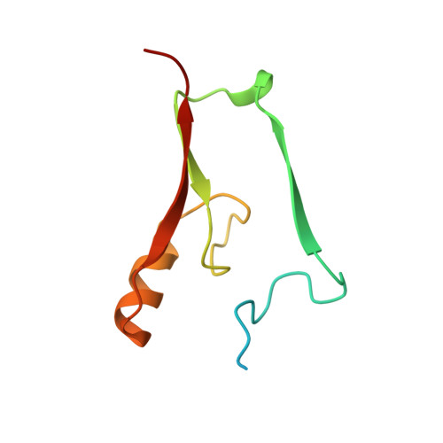
Crystal structure of Miner1: The redox-active 2Fe-2S protein causative in Wolfram Syndrome 2.
Conlan, A.R., Axelrod, H.L., Cohen, A.E., Abresch, E.C., Zuris, J., Yee, D., Nechushtai, R., Jennings, P.A., Paddock, M.L.(2009) J Mol Biol 392: 143-153
- PubMed: 19580816
- DOI: https://doi.org/10.1016/j.jmb.2009.06.079
- Primary Citation of Related Structures:
3FNV - PubMed Abstract:
The endoplasmic reticulum protein Miner1 is essential for health and longevity. Mis-splicing of CISD2, which codes for Miner1, is causative in Wolfram Syndrome 2 (WFS2) resulting in early onset optic atrophy, diabetes mellitus, deafness and decreased lifespan. In knock-out studies, disruption of CISD2 leads to accelerated aging, blindness and muscle atrophy. In this work, we characterized the soluble region of human Miner1 and solved its crystal structure to a resolution of 2.1 A (R-factor=17%). Although originally annotated as a zinc finger, we show that Miner1 is a homodimer harboring two redox-active 2Fe-2S clusters, indicating for the first time an association of a redox-active FeS protein with WFS2. Each 2Fe-2S cluster is bound by a rare Cys(3)-His motif within a 17 amino acid segment. Miner1 is the first functionally different protein that shares the NEET fold with its recently identified paralog mitoNEET, an outer mitochondrial membrane protein. We report the first measurement of the redox potentials (E(m)) of Miner1 and mitoNEET, showing that they are proton-coupled with E(m) approximately 0 mV at pH 7.5. Changes in the pH sensitivity of their cluster stabilities are attributed to significant differences in the electrostatic distribution and surfaces between the two proteins. The structural and biophysical results are discussed in relation to possible roles of Miner1 in cellular Fe-S management and redox reactions.
Organizational Affiliation:
Departments of Chemistry and Biochemistry, University of California at San Diego, La Jolla, 92093, USA.