3FMG

Structure of rotavirus outer capsid protein VP7 trimer in complex with a neutralizing Fab


Experimental Data Snapshot

  • Method: X-RAY DIFFRACTION
  • Resolution: 3.40 Å
  • R-Value Free: 0.238 
  • R-Value Work: 0.224 

wwPDB Validation   3D Report Full Report


This is version 1.2 of the entry. See complete history


Literature

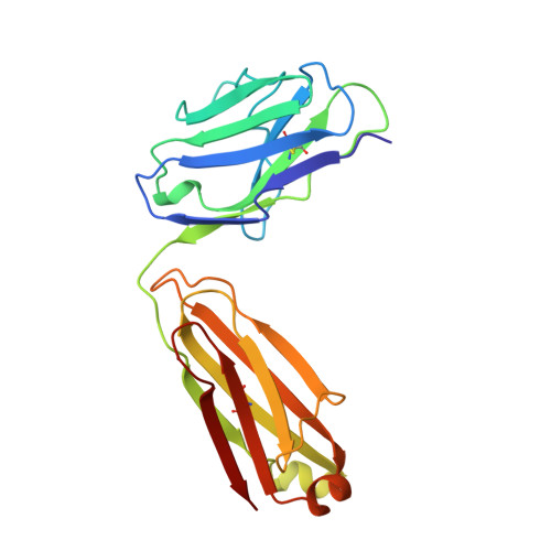
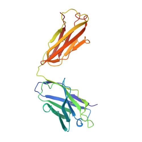
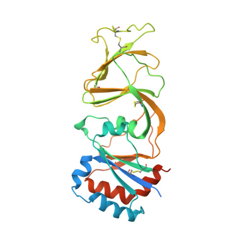
Structure of rotavirus outer-layer protein VP7 bound with a neutralizing Fab.

Aoki, S.T.Settembre, E.C.Trask, S.D.Greenberg, H.B.Harrison, S.C.Dormitzer, P.R.

(2009) Science 324: 1444-1447

  • DOI: https://doi.org/10.1126/science.1170481
  • Primary Citation of Related Structures:  
    3FMG

  • PubMed Abstract: 

    Rotavirus outer-layer protein VP7 is a principal target of protective antibodies. Removal of free calcium ions (Ca2+) dissociates VP7 trimers into monomers, releasing VP7 from the virion, and initiates penetration-inducing conformational changes in the other outer-layer protein, VP4. We report the crystal structure at 3.4 angstrom resolution of VP7 bound with the Fab fragment of a neutralizing monoclonal antibody. The Fab binds across the outer surface of the intersubunit contact, which contains two Ca2+ sites. Mutations that escape neutralization by other antibodies suggest that the same region bears the epitopes of most neutralizing antibodies. The monovalent Fab is sufficient to neutralize infectivity. We propose that neutralizing antibodies against VP7 act by stabilizing the trimer, thereby inhibiting the uncoating trigger for VP4 rearrangement. A disulfide-linked trimer is a potential subunit immunogen.


  • Organizational Affiliation

    Laboratory of Molecular Medicine, Children's Hospital, Boston, MA 02115, USA.


Macromolecules
Find similar proteins by:  (by identity cutoff)  |  3D Structure
Entity ID: 1
MoleculeChains Sequence LengthOrganismDetailsImage
Fab of neutralizing antibody 4F8, light chainA [auth L]211Mus musculusMutation(s): 0 
Entity Groups  
Sequence Clusters30% Identity50% Identity70% Identity90% Identity95% Identity100% Identity
Sequence Annotations
Expand
  • Reference Sequence
Find similar proteins by:  (by identity cutoff)  |  3D Structure
Entity ID: 2
MoleculeChains Sequence LengthOrganismDetailsImage
Fab of neutralizing antibody 4F8, heavy chainB [auth H]221Mus musculusMutation(s): 0 
Entity Groups  
Sequence Clusters30% Identity50% Identity70% Identity90% Identity95% Identity100% Identity
Sequence Annotations
Expand
  • Reference Sequence
Find similar proteins by:  (by identity cutoff)  |  3D Structure
Entity ID: 3
MoleculeChains Sequence LengthOrganismDetailsImage
Glycoprotein VP7C [auth A]276Simian rotavirusMutation(s): 0 
Gene Names: S9VP7
UniProt
Find proteins for P12476 (Rotavirus A (strain RVA/Monkey/United States/RRV/1975/G3P5B[3]))
Explore P12476 
Go to UniProtKB:  P12476
Entity Groups  
Sequence Clusters30% Identity50% Identity70% Identity90% Identity95% Identity100% Identity
UniProt GroupP12476
Sequence Annotations
Expand
  • Reference Sequence
Experimental Data & Validation

Experimental Data

  • Method: X-RAY DIFFRACTION
  • Resolution: 3.40 Å
  • R-Value Free: 0.238 
  • R-Value Work: 0.224 
  • Space Group: P 41 3 2
Unit Cell:
Length ( Å )Angle ( ˚ )
a = 244.179α = 90
b = 244.179β = 90
c = 244.179γ = 90
Software Package:
Software NamePurpose
HKL-2000data collection
CNSrefinement
HKL-2000data reduction
HKL-2000data scaling
CNSphasing

Structure Validation

View Full Validation Report


Entry History 

Deposition Data

Revision History  (Full details and data files)

  • Version 1.0: 2009-06-23
    Type: Initial release
  • Version 1.1: 2011-07-13
    Changes: Version format compliance
  • Version 1.2: 2023-09-06
    Changes: Data collection, Database references, Derived calculations, Refinement description