3FLM

Crystal structure of menD from E.coli


Experimental Data Snapshot

  • Method: X-RAY DIFFRACTION
  • Resolution: 2.70 Å
  • R-Value Free: 0.270 
  • R-Value Work: 0.182 
  • R-Value Observed: 0.186 

wwPDB Validation   3D Report Full Report


This is version 1.4 of the entry. See complete history


Literature

Structural insights of the MenD from Escherichia coli reveal ThDP affinity.

Priyadarshi, A.Saleem, Y.Nam, K.H.Kim, K.S.Park, S.Y.Kim, E.E.Hwang, K.Y.

(2009) Biochem Biophys Res Commun 380: 797-801

  • DOI: https://doi.org/10.1016/j.bbrc.2009.01.168
  • Primary Citation of Related Structures:  
    3FLM

  • PubMed Abstract: 

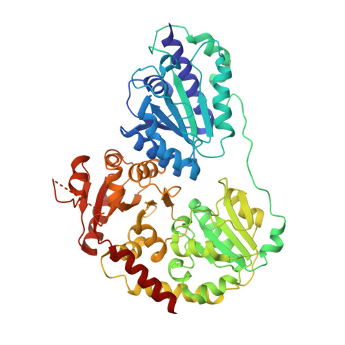
    MenD (2-succinyl-5-enolpyruvyl-6-hydroxy-3-cyclohexadiene-1-carboxylate) synthase belongs to the superfamily of thiamin diphosphate-dependent decarboxylases, which converts isochorismate and 2-oxoglutarate to SHCHC, pyruvate, and carbon dioxide. Here, we report the first crystal structure of apo-MenD from Escherichia coli determined in tetragonal crystal form. The subunit displays the typical three-domain structure observed for ThDP-dependent enzymes. Analytical gel filtration shows that EcMenD behaves as a dimer as well as a tetramer. Circular dichroism and isothermal calorimetry results confirm EcMenD dependency on ThDP, which concomitantly helps to stabilize with better configuration.


  • Organizational Affiliation

    Biomedical Research Center, Life Science Division, Korea Institute of Science and Technology, Seongbuk-gu, Seoul 136-791, Republic of Korea.


Macromolecules
Find similar proteins by:  (by identity cutoff)  |  3D Structure
Entity ID: 1
MoleculeChains Sequence LengthOrganismDetailsImage
2-succinyl-5-enolpyruvyl-6-hydroxy-3-cyclohexene-1-carboxylate synthase
A, B
556Escherichia coli K-12Mutation(s): 1 
Gene Names: b2264JW5374menD
EC: 2.2.1.9
UniProt
Find proteins for P17109 (Escherichia coli (strain K12))
Explore P17109 
Go to UniProtKB:  P17109
Entity Groups  
Sequence Clusters30% Identity50% Identity70% Identity90% Identity95% Identity100% Identity
UniProt GroupP17109
Sequence Annotations
Expand
  • Reference Sequence
Experimental Data & Validation

Experimental Data

  • Method: X-RAY DIFFRACTION
  • Resolution: 2.70 Å
  • R-Value Free: 0.270 
  • R-Value Work: 0.182 
  • R-Value Observed: 0.186 
  • Space Group: P 41 21 2
Unit Cell:
Length ( Å )Angle ( ˚ )
a = 118.067α = 90
b = 118.067β = 90
c = 176.477γ = 90
Software Package:
Software NamePurpose
HKL-2000data collection
MOLREPphasing
REFMACrefinement
HKL-2000data reduction
HKL-2000data scaling

Structure Validation

View Full Validation Report


Entry History 

Deposition Data

Revision History  (Full details and data files)

  • Version 1.0: 2009-03-24
    Type: Initial release
  • Version 1.1: 2011-07-13
    Changes: Version format compliance
  • Version 1.2: 2012-04-18
    Changes: Database references
  • Version 1.3: 2021-11-10
    Changes: Database references
  • Version 1.4: 2023-11-01
    Changes: Data collection, Refinement description