3FE4

Crystal Structure of Human Carbonic Anhydrase vi


Experimental Data Snapshot

  • Method: X-RAY DIFFRACTION
  • Resolution: 1.90 Å
  • R-Value Free: 0.235 
  • R-Value Work: 0.177 
  • R-Value Observed: 0.179 

wwPDB Validation   3D Report Full Report


This is version 1.4 of the entry. See complete history


Literature

Crystal structure of the secretory isozyme of mammalian carbonic anhydrases CA VI: implications for biological assembly and inhibitor development

Pilka, E.S.Kochan, G.Oppermann, U.Yue, W.W.

(2012) Biochem Biophys Res Commun 419: 485-489

  • DOI: https://doi.org/10.1016/j.bbrc.2012.02.038
  • Primary Citation of Related Structures:  
    3FE4

  • PubMed Abstract: 

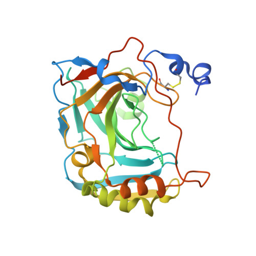
    Zn(2+)-dependent carbonic anhydrases (CA) catalyse the reversible hydration of carbon dioxide to bicarbonate and participate in diverse physiological processes, hence having manifold therapeutic potentials. Among the 15 human CAs with wide-ranging sub-cellular localisation and kinetic properties, CA VI is the only secretory isoform. The 1.9Å crystal structure of the human CA VI catalytic domain reveals a prototypical mammalian CA fold, and a novel dimeric arrangement as compared to previously-reported CA structures. The active site cavity contains a cluster of non-conserved residues that may be involved in ligand binding and have significant implications for developing the next-generation of isoform-specific inhibitors.


  • Organizational Affiliation

    Structural Genomics Consortium, Oxford, UK.


Macromolecules
Find similar proteins by:  (by identity cutoff)  |  3D Structure
Entity ID: 1
MoleculeChains Sequence LengthOrganismDetailsImage
Carbonic anhydrase 6
A, B
278Homo sapiensMutation(s): 0 
Gene Names: CA6
EC: 4.2.1.1
UniProt & NIH Common Fund Data Resources
Find proteins for P23280 (Homo sapiens)
Explore P23280 
Go to UniProtKB:  P23280
PHAROS:  P23280
GTEx:  ENSG00000131686 
Entity Groups  
Sequence Clusters30% Identity50% Identity70% Identity90% Identity95% Identity100% Identity
UniProt GroupP23280
Sequence Annotations
Expand
  • Reference Sequence
Experimental Data & Validation

Experimental Data

  • Method: X-RAY DIFFRACTION
  • Resolution: 1.90 Å
  • R-Value Free: 0.235 
  • R-Value Work: 0.177 
  • R-Value Observed: 0.179 
  • Space Group: P 21 21 21
Unit Cell:
Length ( Å )Angle ( ˚ )
a = 51.65α = 90
b = 80.048β = 90
c = 133.441γ = 90
Software Package:
Software NamePurpose
PHASERphasing
REFMACrefinement
MOSFLMdata reduction
SCALAdata scaling

Structure Validation

View Full Validation Report


Entry History 

Revision History  (Full details and data files)

  • Version 1.0: 2008-12-16
    Type: Initial release
  • Version 1.1: 2011-07-13
    Changes: Advisory, Version format compliance
  • Version 1.2: 2013-11-27
    Changes: Database references
  • Version 1.3: 2017-11-01
    Changes: Refinement description
  • Version 1.4: 2023-11-01
    Changes: Data collection, Database references, Derived calculations, Refinement description