3EYO

Crystal structure of anti-human cytomegalovirus antibody 8F9


Experimental Data Snapshot

  • Method: X-RAY DIFFRACTION
  • Resolution: 2.50 Å
  • R-Value Free: 0.268 
  • R-Value Work: 0.241 
  • R-Value Observed: 0.241 

wwPDB Validation   3D Report Full Report


This is version 1.2 of the entry. See complete history


Literature

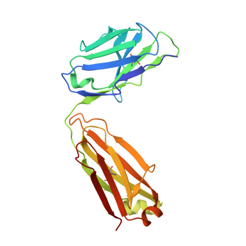
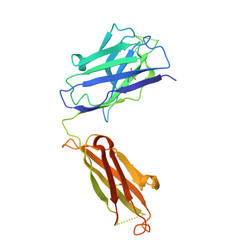
Germline V-genes sculpt the binding site of a family of antibodies neutralizing human cytomegalovirus.

Thomson, C.A.Bryson, S.McLean, G.R.Creagh, A.L.Pai, E.F.Schrader, J.W.

(2008) EMBO J 27: 2592-2602

  • DOI: https://doi.org/10.1038/emboj.2008.179
  • Primary Citation of Related Structures:  
    3EYF, 3EYO, 3EYQ, 3F12

  • PubMed Abstract: 

    Immunoglobulin genes are generated somatically through specialized mechanisms resulting in a vast repertoire of antigen-binding sites. Despite the stochastic nature of these processes, the V-genes that encode most of the antigen-combining site are under positive evolutionary selection, raising the possibility that V-genes have been selected to encode key structural features of binding sites of protective antibodies against certain pathogens. Human, neutralizing antibodies to human cytomegalovirus that bind the AD-2S1 epitope on its gB envelope protein repeatedly use a pair of well-conserved, germline V-genes IGHV3-30 and IGKV3-11. Here, we present crystallographic, kinetic and thermodynamic analyses of the binding site of such an antibody and that of its primary immunoglobulin ancestor. These show that these germline V-genes encode key side chain contacts with the viral antigen and thereby dictate key structural features of the hypermutated, high-affinity neutralizing antibody. V-genes may thus encode an innate, protective immunological memory that targets vulnerable, invariant sites on multiple pathogens.


  • Organizational Affiliation

    The Biomedical Research Centre, University of British Columbia, Vancouver, British Columbia, Canada.


Macromolecules
Find similar proteins by:  (by identity cutoff)  |  3D Structure
Entity ID: 1
MoleculeChains Sequence LengthOrganismDetailsImage
8f9 Fab
A, C
216Homo sapiensMutation(s): 0 
UniProt
Find proteins for Q6PJF2 (Homo sapiens)
Explore Q6PJF2 
Go to UniProtKB:  Q6PJF2
Entity Groups  
Sequence Clusters30% Identity50% Identity70% Identity90% Identity95% Identity100% Identity
UniProt GroupQ6PJF2
Sequence Annotations
Expand
  • Reference Sequence
Find similar proteins by:  (by identity cutoff)  |  3D Structure
Entity ID: 2
MoleculeChains Sequence LengthOrganismDetailsImage
AD-2
B, D
242Homo sapiensMutation(s): 0 
UniProt
Find proteins for Q0ZCG9 (Homo sapiens)
Explore Q0ZCG9 
Go to UniProtKB:  Q0ZCG9
Entity Groups  
Sequence Clusters30% Identity50% Identity70% Identity90% Identity95% Identity100% Identity
UniProt GroupQ0ZCG9
Sequence Annotations
Expand
  • Reference Sequence
Experimental Data & Validation

Experimental Data

  • Method: X-RAY DIFFRACTION
  • Resolution: 2.50 Å
  • R-Value Free: 0.268 
  • R-Value Work: 0.241 
  • R-Value Observed: 0.241 
  • Space Group: P 21 21 21
Unit Cell:
Length ( Å )Angle ( ˚ )
a = 63.03α = 90
b = 110.73β = 90
c = 180.25γ = 90
Software Package:
Software NamePurpose
HKL-2000data collection
CNSrefinement
HKL-2000data reduction
HKL-2000data scaling
CNSphasing

Structure Validation

View Full Validation Report


Entry History 

Deposition Data

Revision History  (Full details and data files)

  • Version 1.0: 2008-12-16
    Type: Initial release
  • Version 1.1: 2011-07-13
    Changes: Version format compliance
  • Version 1.2: 2023-09-06
    Changes: Data collection, Database references, Refinement description