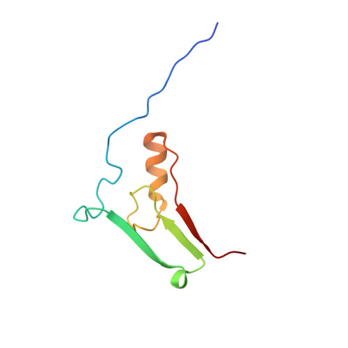
The novel 2Fe-2S outer mitochondrial protein mitoNEET displays conformational flexibility in its N-terminal cytoplasmic tethering domain.
Conlan, A.R., Paddock, M.L., Axelrod, H.L., Cohen, A.E., Abresch, E.C., Wiley, S., Roy, M., Nechushtai, R., Jennings, P.A.(2009) Acta Crystallogr Sect F Struct Biol Cryst Commun 65: 654-659
- PubMed: 19574633
- DOI: https://doi.org/10.1107/S1744309109019605
- Primary Citation of Related Structures:
3EW0 - PubMed Abstract:
A primary role for mitochondrial dysfunction is indicated in the pathogenesis of insulin resistance. A widely used drug for the treatment of type 2 diabetes is pioglitazone, a member of the thiazolidinedione class of molecules. MitoNEET, a 2Fe-2S outer mitochondrial membrane protein, binds pioglitazone [Colca et al. (2004), Am. J. Physiol. Endocrinol. Metab. 286, E252-E260]. The soluble domain of the human mitoNEET protein has been expressed C-terminal to the superfolder green fluorescent protein and the mitoNEET protein has been isolated. Comparison of the crystal structure of mitoNEET isolated from cleavage of the fusion protein (1.4 A resolution, R factor = 20.2%) with other solved structures shows that the CDGSH domains are superimposable, indicating proper assembly of mitoNEET. Furthermore, there is considerable flexibility in the position of the cytoplasmic tethering arms, resulting in two different conformations in the crystal structure. This flexibility affords multiple orientations on the outer mitochondrial membrane.
Organizational Affiliation:
Department of Chemistry, University of California, San Diego, La Jolla, CA 92093, USA.