3ETC

2.1 A structure of acyl-adenylate synthetase from Methanosarcina acetivorans containing a link between Lys256 and Cys298


Experimental Data Snapshot

  • Method: X-RAY DIFFRACTION
  • Resolution: 2.10 Å
  • R-Value Free: 0.207 
  • R-Value Work: 0.175 
  • R-Value Observed: 0.177 

wwPDB Validation   3D Report Full Report


This is version 1.2 of the entry. See complete history


Literature

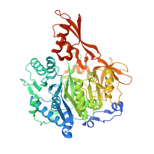
The 2.1 A crystal structure of an acyl-CoA synthetase from Methanosarcina acetivorans reveals an alternate acyl-binding pocket for small branched acyl substrates.

Shah, M.B.Ingram-Smith, C.Cooper, L.L.Qu, J.Meng, Y.Smith, K.S.Gulick, A.M.

(2009) Proteins 77: 685-698

  • DOI: https://doi.org/10.1002/prot.22482
  • Primary Citation of Related Structures:  
    3ETC

  • PubMed Abstract: 

    The acyl-AMP forming family of adenylating enzymes catalyze two-step reactions to activate a carboxylate with the chemical energy derived from ATP hydrolysis. X-ray crystal structures have been determined for multiple members of this family and, together with biochemical studies, provide insights into the active site and catalytic mechanisms used by these enzymes. These studies have shown that the enzymes use a domain rotation of 140 degrees to reconfigure a single active site to catalyze the two partial reactions. We present here the crystal structure of a new medium chain acyl-CoA synthetase from Methanosarcina acetivorans. The binding pocket for the three substrates is analyzed, with many conserved residues present in the AMP binding pocket. The CoA binding pocket is compared to the pockets of both acetyl-CoA synthetase and 4-chlorobenzoate:CoA ligase. Most interestingly, the acyl-binding pocket of the new structure is compared with other acyl- and aryl-CoA synthetases. A comparison of the acyl-binding pocket of the acyl-CoA synthetase from M. acetivorans with other structures identifies a shallow pocket that is used to bind the medium chain carboxylates. These insights emphasize the high sequence and structural diversity among this family in the area of the acyl-binding pocket.


  • Organizational Affiliation

    Hauptman-Woodward Medical Research Institute, Buffalo, New York 14203-1102, USA.


Macromolecules
Find similar proteins by:  (by identity cutoff)  |  3D Structure
Entity ID: 1
MoleculeChains Sequence LengthOrganismDetailsImage
AMP-binding protein
A, B
580Methanosarcina acetivoransMutation(s): 0 
Gene Names: MA_2912Methanosarcina Acetivorans
EC: 6.2.1
UniProt
Find proteins for Q8TLW1 (Methanosarcina acetivorans (strain ATCC 35395 / DSM 2834 / JCM 12185 / C2A))
Explore Q8TLW1 
Go to UniProtKB:  Q8TLW1
Entity Groups  
Sequence Clusters30% Identity50% Identity70% Identity90% Identity95% Identity100% Identity
UniProt GroupQ8TLW1
Sequence Annotations
Expand
  • Reference Sequence
Small Molecules
Ligands 7 Unique
IDChains Name / Formula / InChI Key2D Diagram3D Interactions
EPE
Query on EPE

Download Ideal Coordinates CCD File 
G [auth A],
H [auth A],
R [auth B]
4-(2-HYDROXYETHYL)-1-PIPERAZINE ETHANESULFONIC ACID
C8 H18 N2 O4 S
JKMHFZQWWAIEOD-UHFFFAOYSA-N
1PE
Query on 1PE

Download Ideal Coordinates CCD File 
E [auth A],
F [auth A]
PENTAETHYLENE GLYCOL
C10 H22 O6
JLFNLZLINWHATN-UHFFFAOYSA-N
PGE
Query on PGE

Download Ideal Coordinates CCD File 
D [auth A],
P [auth B],
Q [auth B]
TRIETHYLENE GLYCOL
C6 H14 O4
ZIBGPFATKBEMQZ-UHFFFAOYSA-N
GOL
Query on GOL

Download Ideal Coordinates CCD File 
K [auth A]
L [auth A]
M [auth A]
N [auth A]
U [auth B]
K [auth A],
L [auth A],
M [auth A],
N [auth A],
U [auth B],
V [auth B],
W [auth B]
GLYCEROL
C3 H8 O3
PEDCQBHIVMGVHV-UHFFFAOYSA-N
NO3
Query on NO3

Download Ideal Coordinates CCD File 
J [auth A],
T [auth B]
NITRATE ION
N O3
NHNBFGGVMKEFGY-UHFFFAOYSA-N
FOR
Query on FOR

Download Ideal Coordinates CCD File 
C [auth A],
O [auth B]
FORMYL GROUP
C H2 O
WSFSSNUMVMOOMR-UHFFFAOYSA-N
MG
Query on MG

Download Ideal Coordinates CCD File 
I [auth A],
S [auth B]
MAGNESIUM ION
Mg
JLVVSXFLKOJNIY-UHFFFAOYSA-N
Experimental Data & Validation

Experimental Data

  • Method: X-RAY DIFFRACTION
  • Resolution: 2.10 Å
  • R-Value Free: 0.207 
  • R-Value Work: 0.175 
  • R-Value Observed: 0.177 
  • Space Group: P 21 21 21
Unit Cell:
Length ( Å )Angle ( ˚ )
a = 91.699α = 90
b = 96.371β = 90
c = 141.543γ = 90
Software Package:
Software NamePurpose
Blu-Icedata collection
SnBphasing
REFMACrefinement
HKL-2000data reduction
HKL-2000data scaling

Structure Validation

View Full Validation Report


Entry History 

Deposition Data

Revision History  (Full details and data files)

  • Version 1.0: 2009-07-07
    Type: Initial release
  • Version 1.1: 2011-07-13
    Changes: Advisory, Version format compliance
  • Version 1.2: 2023-12-27
    Changes: Data collection, Database references, Derived calculations