3EOY

Structure of Reovirus sigma1 in Complex with Its Receptor Junctional Adhesion Molecule-A


Experimental Data Snapshot

  • Method: X-RAY DIFFRACTION
  • Resolution: 3.40 Å
  • R-Value Free: 0.252 
  • R-Value Work: 0.210 
  • R-Value Observed: 0.210 

wwPDB Validation   3D Report Full Report


This is version 1.3 of the entry. See complete history


Literature

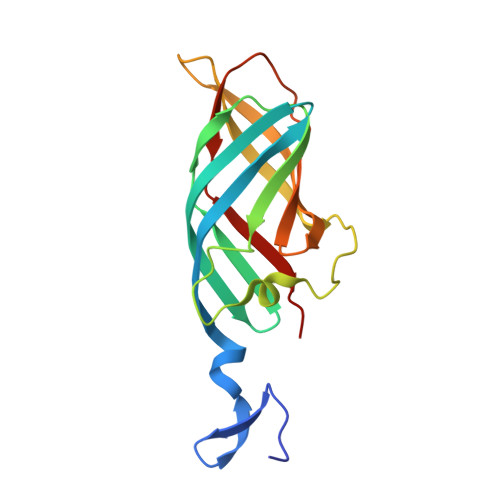
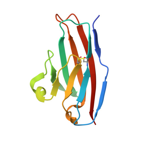
Structure of reovirus sigma1 in complex with its receptor junctional adhesion molecule-A

Kirchner, E.Guglielmi, K.M.Strauss, H.M.Dermody, T.S.Stehle, T.

(2008) PLoS Pathog 4: e1000235-e1000235

  • DOI: https://doi.org/10.1371/journal.ppat.1000235
  • Primary Citation of Related Structures:  
    3EOY

  • PubMed Abstract: 

    Viral attachment to specific host receptors is the first step in viral infection and serves an essential function in the selection of target cells. Mammalian reoviruses are highly useful experimental models for studies of viral pathogenesis and show promise as vectors for oncolytics and vaccines. Reoviruses engage cells by binding to carbohydrates and the immunoglobulin superfamily member, junctional adhesion molecule-A (JAM-A). JAM-A exists at the cell surface as a homodimer formed by extensive contacts between its N-terminal immunoglobulin-like domains. We report the crystal structure of reovirus attachment protein sigma1 in complex with a soluble form of JAM-A. The sigma1 protein disrupts the JAM-A dimer, engaging a single JAM-A molecule via virtually the same interface that is used for JAM-A homodimerization. Thus, reovirus takes advantage of the adhesive nature of an immunoglobulin-superfamily receptor by usurping the ligand-binding site of this molecule to attach to the cell surface. The dissociation constant (K(D)) of the interaction between sigma1 and JAM-A is 1,000-fold lower than that of the homophilic interaction between JAM-A molecules, indicating that JAM-A strongly prefers sigma1 as a ligand. Analysis of reovirus mutants engineered by plasmid-based reverse genetics revealed residues in sigma1 required for binding to JAM-A and infectivity of cultured cells. These studies define biophysical mechanisms of reovirus cell attachment and provide a platform for manipulating reovirus tropism to enhance vector targeting.


  • Organizational Affiliation

    Interfaculty Institute for Biochemistry, University of Tuebingen, Tuebingen, Germany.


Macromolecules
Find similar proteins by:  (by identity cutoff)  |  3D Structure
Entity ID: 1
MoleculeChains Sequence LengthOrganismDetailsImage
Outer capsid protein sigma-1
A, B, C, D, E
A, B, C, D, E, F
165Mammalian orthoreovirus 3 DearingMutation(s): 0 
Gene Names: S1
UniProt
Find proteins for P03528 (Reovirus type 3 (strain Dearing))
Explore P03528 
Go to UniProtKB:  P03528
Entity Groups  
Sequence Clusters30% Identity50% Identity70% Identity90% Identity95% Identity100% Identity
UniProt GroupP03528
Sequence Annotations
Expand
  • Reference Sequence
Find similar proteins by:  (by identity cutoff)  |  3D Structure
Entity ID: 2
MoleculeChains Sequence LengthOrganismDetailsImage
Junctional adhesion molecule A
G, H, I, J, K
G, H, I, J, K, L
104Homo sapiensMutation(s): 0 
UniProt & NIH Common Fund Data Resources
Find proteins for Q9Y624 (Homo sapiens)
Explore Q9Y624 
Go to UniProtKB:  Q9Y624
PHAROS:  Q9Y624
GTEx:  ENSG00000158769 
Entity Groups  
Sequence Clusters30% Identity50% Identity70% Identity90% Identity95% Identity100% Identity
UniProt GroupQ9Y624
Sequence Annotations
Expand
  • Reference Sequence
Experimental Data & Validation

Experimental Data

  • Method: X-RAY DIFFRACTION
  • Resolution: 3.40 Å
  • R-Value Free: 0.252 
  • R-Value Work: 0.210 
  • R-Value Observed: 0.210 
  • Space Group: P 21 21 2
Unit Cell:
Length ( Å )Angle ( ˚ )
a = 105.94α = 90
b = 124.34β = 90
c = 130.659γ = 90
Software Package:
Software NamePurpose
DENZOdata reduction
SCALEPACKdata scaling
PHASERphasing
CNSrefinement
PDB_EXTRACTdata extraction
MAR345data collection

Structure Validation

View Full Validation Report


Entry History 

Deposition Data

Revision History  (Full details and data files)

  • Version 1.0: 2008-11-04
    Type: Initial release
  • Version 1.1: 2011-07-13
    Changes: Version format compliance
  • Version 1.2: 2017-10-25
    Changes: Refinement description
  • Version 1.3: 2023-11-01
    Changes: Data collection, Database references, Refinement description