


2.5A crystal structure of glucose/ribitol dehydrogenase from brucella melitensis
Seattle Structural Genomics Center for Infectious Disease (SSGCID)To be published.
Experimental Data Snapshot
wwPDB Validation 3D Report Full Report

Entity ID: 1 | |||||
|---|---|---|---|---|---|
| Molecule | Chains | Sequence Length | Organism | Details | Image |
| GLUCOSE/RIBITOL DEHYDROGENASE | 246 | Brucella melitensis | Mutation(s): 0 Gene Names: BMEI1477 | 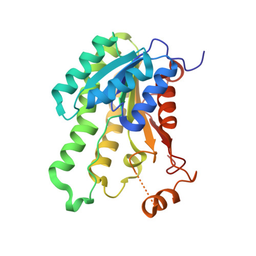 | |
UniProt | |||||
Find proteins for Q2YMG6 (Brucella abortus (strain 2308)) Explore Q2YMG6 Go to UniProtKB: Q2YMG6 | |||||
Entity Groups | |||||
| Sequence Clusters | 30% Identity50% Identity70% Identity90% Identity95% Identity100% Identity | ||||
| UniProt Group | Q2YMG6 | ||||
Sequence AnnotationsExpand | |||||
| |||||
| Length ( Å ) | Angle ( ˚ ) |
|---|---|
| a = 99.489 | α = 90 |
| b = 147.939 | β = 90 |
| c = 63.892 | γ = 90 |
| Software Name | Purpose |
|---|---|
| DENZO | data reduction |
| SCALEPACK | data scaling |
| PHASER | phasing |
| REFMAC | refinement |
| PDB_EXTRACT | data extraction |
RCSB PDB (citation) is hosted by
RCSB PDB is a member of the

