3ECB

Crystal structure of mouse H-2Dd in complex with peptide P18-I10 derived from human immunodeficiency virus envelope glycoprotein 120


Experimental Data Snapshot

  • Method: X-RAY DIFFRACTION
  • Resolution: 1.70 Å
  • R-Value Free: 0.227 
  • R-Value Work: 0.182 
  • R-Value Observed: 0.184 

wwPDB Validation   3D Report Full Report


This is version 1.2 of the entry. See complete history


Literature

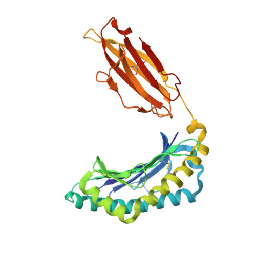
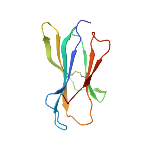
Structural basis of the CD8alphabeta/MHC class i interaction: focused recognition orients CD8beta to a T cell proximal position

Wang, R.Natarajan, K.Margulies, D.H.

(2009) J Immunol 183: 2554-2564

  • DOI: https://doi.org/10.4049/jimmunol.0901276
  • Primary Citation of Related Structures:  
    3DMM, 3ECB

  • PubMed Abstract: 

    In the immune system, B cells, dendritic cells, NK cells, and T lymphocytes all respond to signals received via ligand binding to receptors and coreceptors. Although the specificity of T cell recognition is determined by the interaction of T cell receptors with MHC/peptide complexes, the development of T cells in the thymus and their sensitivity to Ag are also dependent on coreceptor molecules CD8 (for MHC class I (MHCI)) and CD4 (for MHCII). The CD8alphabeta heterodimer is a potent coreceptor for T cell activation, but efforts to understand its function fully have been hampered by ignorance of the structural details of its interactions with MHCI. In this study we describe the structure of CD8alphabeta in complex with the murine MHCI molecule H-2D(d) at 2.6 A resolution. The focus of the CD8alphabeta interaction is the acidic loop (residues 222-228) of the alpha3 domain of H-2D(d). The beta subunit occupies a T cell membrane proximal position, defining the relative positions of the CD8alpha and CD8beta subunits. Unlike the CD8alphaalpha homodimer, CD8alphabeta does not contact the MHCI alpha(2)- or beta(2)-microglobulin domains. Movements of the CD8alpha CDR2 and CD8beta CDR1 and CDR2 loops as well as the flexibility of the H-2D(d) CD loop facilitate the monovalent interaction. The structure resolves inconclusive data on the topology of the CD8alphabeta/MHCI interaction, indicates that CD8beta is crucial in orienting the CD8alphabeta heterodimer, provides a framework for understanding the mechanistic role of CD8alphabeta in lymphoid cell signaling, and offers a tangible context for design of structurally altered coreceptors for tumor and viral immunotherapy.


  • Organizational Affiliation

    Molecular Biology Section, Laboratory of Immunology, National Institute of Allergy and Infectious Diseases, National Institutes of Health, Bethesda, MD 20892, USA.


Macromolecules
Find similar proteins by:  (by identity cutoff)  |  3D Structure
Entity ID: 1
MoleculeChains Sequence LengthOrganismDetailsImage
H-2 class I histocompatibility antigen, D-D alpha chain277Mus musculusMutation(s): 0 
Gene Names: H2-D1
UniProt
Find proteins for P01900 (Mus musculus)
Explore P01900 
Go to UniProtKB:  P01900
Entity Groups  
Sequence Clusters30% Identity50% Identity70% Identity90% Identity95% Identity100% Identity
UniProt GroupP01900
Sequence Annotations
Expand
  • Reference Sequence
Find similar proteins by:  (by identity cutoff)  |  3D Structure
Entity ID: 2
MoleculeChains Sequence LengthOrganismDetailsImage
Beta-2 microglobulin100Mus musculusMutation(s): 0 
Gene Names: B2mRP23-34E24.5-001
UniProt & NIH Common Fund Data Resources
Find proteins for P01887 (Mus musculus)
Explore P01887 
Go to UniProtKB:  P01887
IMPC:  MGI:88127
Entity Groups  
Sequence Clusters30% Identity50% Identity70% Identity90% Identity95% Identity100% Identity
UniProt GroupP01887
Sequence Annotations
Expand
  • Reference Sequence

Find similar proteins by:  Sequence   |   3D Structure  

Entity ID: 3
MoleculeChains Sequence LengthOrganismDetailsImage
Peptide P18-I10 from HIV gp160C [auth P]10N/AMutation(s): 0 
UniProt
Find proteins for P19551 (Human immunodeficiency virus type 1 group M subtype B (isolate MFA))
Explore P19551 
Go to UniProtKB:  P19551
Entity Groups  
Sequence Clusters30% Identity50% Identity70% Identity90% Identity95% Identity100% Identity
UniProt GroupP19551
Sequence Annotations
Expand
  • Reference Sequence
Experimental Data & Validation

Experimental Data

  • Method: X-RAY DIFFRACTION
  • Resolution: 1.70 Å
  • R-Value Free: 0.227 
  • R-Value Work: 0.182 
  • R-Value Observed: 0.184 
  • Space Group: P 21 21 21
Unit Cell:
Length ( Å )Angle ( ˚ )
a = 46.727α = 90
b = 89.452β = 90
c = 109.705γ = 90
Software Package:
Software NamePurpose
ADSCdata collection
AMoREphasing
PHENIXrefinement
HKL-2000data reduction
SCALEPACKdata scaling

Structure Validation

View Full Validation Report


Entry History 

Deposition Data

Revision History  (Full details and data files)

  • Version 1.0: 2009-07-14
    Type: Initial release
  • Version 1.1: 2011-07-13
    Changes: Version format compliance
  • Version 1.2: 2023-08-30
    Changes: Data collection, Database references, Derived calculations, Refinement description