3E37

Protein farnesyltransferase complexed with bisubstrate ethylenediamine scaffold inhibitor 5


Experimental Data Snapshot

  • Method: X-RAY DIFFRACTION
  • Resolution: 1.80 Å
  • R-Value Free: 0.190 
  • R-Value Work: 0.168 
  • R-Value Observed: 0.169 

wwPDB Validation   3D Report Full Report


Ligand Structure Quality Assessment 


This is version 2.1 of the entry. See complete history


Literature

Structural basis for binding and selectivity of antimalarial and anticancer ethylenediamine inhibitors to protein farnesyltransferase.

Hast, M.A.Fletcher, S.Cummings, C.G.Pusateri, E.E.Blaskovich, M.A.Rivas, K.Gelb, M.H.Van Voorhis, W.C.Sebti, S.M.Hamilton, A.D.Beese, L.S.

(2009) Chem Biol 16: 181-192

  • DOI: https://doi.org/10.1016/j.chembiol.2009.01.014
  • Primary Citation of Related Structures:  
    3E30, 3E32, 3E33, 3E34, 3E37

  • PubMed Abstract: 

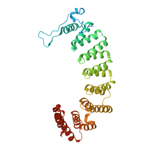
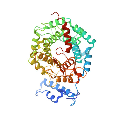
    Protein farnesyltransferase (FTase) catalyzes an essential posttranslational lipid modification of more than 60 proteins involved in intracellular signal transduction networks. FTase inhibitors have emerged as a significant target for development of anticancer therapeutics and, more recently, for the treatment of parasitic diseases caused by protozoan pathogens, including malaria (Plasmodium falciparum). We present the X-ray crystallographic structures of complexes of mammalian FTase with five inhibitors based on an ethylenediamine scaffold, two of which exhibit over 1000-fold selective inhibition of P. falciparum FTase. These structures reveal the dominant determinants in both the inhibitor and enzyme that control binding and selectivity. Comparison to a homology model constructed for the P. falciparum FTase suggests opportunities for further improving selectivity of a new generation of antimalarial inhibitors.


  • Organizational Affiliation

    Department of Biochemistry, Duke University Medical Center, Box 3711, Durham, NC 27710, USA.


Macromolecules
Find similar proteins by:  (by identity cutoff)  |  3D Structure
Entity ID: 1
MoleculeChains Sequence LengthOrganismDetailsImage
Protein farnesyltransferase/geranylgeranyltransferase type-1 subunit alpha379Homo sapiensMutation(s): 0 
Gene Names: FNTA
EC: 2.5.1.58 (PDB Primary Data), 2.5.1.59 (PDB Primary Data)
UniProt & NIH Common Fund Data Resources
Find proteins for P49354 (Homo sapiens)
Explore P49354 
Go to UniProtKB:  P49354
PHAROS:  P49354
GTEx:  ENSG00000168522 
Entity Groups  
Sequence Clusters30% Identity50% Identity70% Identity90% Identity95% Identity100% Identity
UniProt GroupP49354
Sequence Annotations
Expand
  • Reference Sequence
Find similar proteins by:  (by identity cutoff)  |  3D Structure
Entity ID: 2
MoleculeChains Sequence LengthOrganismDetailsImage
Protein farnesyltransferase subunit beta437Homo sapiensMutation(s): 0 
Gene Names: FNTB
EC: 2.5.1.58
UniProt & NIH Common Fund Data Resources
Find proteins for P49356 (Homo sapiens)
Explore P49356 
Go to UniProtKB:  P49356
PHAROS:  P49356
GTEx:  ENSG00000257365 
Entity Groups  
Sequence Clusters30% Identity50% Identity70% Identity90% Identity95% Identity100% Identity
UniProt GroupP49356
Sequence Annotations
Expand
  • Reference Sequence
Oligosaccharides

Help

Entity ID: 3
MoleculeChains Length2D Diagram Glycosylation3D Interactions
beta-D-fructofuranose-(2-1)-alpha-D-glucopyranose
C
2N/A
Glycosylation Resources
GlyTouCan:  G05551OP
GlyCosmos:  G05551OP
Small Molecules
Ligands 2 Unique
IDChains Name / Formula / InChI Key2D Diagram3D Interactions
ED5
Query on ED5

Download Ideal Coordinates CCD File 
D [auth B]tert-butyl 4-({(2-{(4-cyanophenyl)[(1-methyl-1H-imidazol-5-yl)methyl]amino}ethyl)[(2-methylphenyl)sulfonyl]amino}methyl)piperidine-1-carboxylate
C32 H42 N6 O4 S
CHEKEDNUVFBGCF-UHFFFAOYSA-N
ZN
Query on ZN

Download Ideal Coordinates CCD File 
E [auth B]ZINC ION
Zn
PTFCDOFLOPIGGS-UHFFFAOYSA-N
Binding Affinity Annotations 
IDSourceBinding Affinity
ED5 BindingDB:  3E37 IC50: 4050 (nM) from 1 assay(s)
Binding MOAD:  3E37 IC50: 4050 (nM) from 1 assay(s)
Biologically Interesting Molecules (External Reference) 1 Unique
Experimental Data & Validation

Experimental Data

  • Method: X-RAY DIFFRACTION
  • Resolution: 1.80 Å
  • R-Value Free: 0.190 
  • R-Value Work: 0.168 
  • R-Value Observed: 0.169 
  • Space Group: P 61
Unit Cell:
Length ( Å )Angle ( ˚ )
a = 178.386α = 90
b = 178.386β = 90
c = 64.51γ = 120
Software Package:
Software NamePurpose
REFMACrefinement
HKL-2000data collection
HKL-2000data reduction
HKL-2000data scaling
PHASERphasing

Structure Validation

View Full Validation Report


Ligand Structure Quality Assessment 


Entry History 

Deposition Data

Revision History  (Full details and data files)

  • Version 1.0: 2009-03-10
    Type: Initial release
  • Version 1.1: 2011-07-13
    Changes: Version format compliance
  • Version 2.0: 2020-07-29
    Type: Remediation
    Reason: Carbohydrate remediation
    Changes: Atomic model, Data collection, Derived calculations, Non-polymer description, Structure summary
  • Version 2.1: 2024-02-21
    Changes: Data collection, Database references, Structure summary