3E2B

Crystal structure of Dynein Light chain LC8 in complex with a peptide derived from Swallow


Experimental Data Snapshot

  • Method: X-RAY DIFFRACTION
  • Resolution: 2.00 Å
  • R-Value Free: 0.266 
  • R-Value Work: 0.216 
  • R-Value Observed: 0.221 

wwPDB Validation   3D Report Full Report


This is version 1.2 of the entry. See complete history


Literature

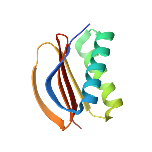
The interplay of ligand binding and quaternary structure in the diverse interactions of dynein light chain LC8.

Benison, G.Karplus, P.A.Barbar, E.

(2008) J Mol Biol 384: 954-966

  • DOI: https://doi.org/10.1016/j.jmb.2008.09.083
  • Primary Citation of Related Structures:  
    3BRI, 3E2B

  • PubMed Abstract: 

    Dynein light chain LC8 is a small, dimeric, and very highly conserved globular protein that is an integral part of the dynein and myosin molecular motors but appears to have a broader role in multiple protein complexes unrelated to molecular motors. LC8 binds to two families of targets: those having a KXTQT sequence fingerprint and those having a GIQVD fingerprint. All known LC8 binding partners containing these fingerprints share a common binding site on LC8 that raises the question of what determines binding specificity. Here, we present the crystal structure of apo-LC8 at 1.7-A resolution, which, when compared with the crystal structures of several LC8 complexes, gives insight into the mechanism underlying the binding diversity of LC8. Peptide binding is associated with a shift in quaternary structure that expands the hydrophobic binding surface available to the ligand, in addition to changes in tertiary structure and ordering of LC8 around the binding groove. The observed quaternary shift suggests a mechanism by which binding at one of the two identical sites can influence binding at the other. NMR spectra of titrations with peptides from each fingerprint family show evidence of allosteric interaction between the two binding sites, to a differing degree in the two ligand families. Allosteric interaction between the binding sites may be a mechanism to promote simultaneous binding of ligands from the same family, providing a physiological role for the two fingerprints.


  • Organizational Affiliation

    Department of Biochemistry and Biophysics, Oregon State University, Corvallis, OR 97331, USA.


Macromolecules
Find similar proteins by:  (by identity cutoff)  |  3D Structure
Entity ID: 1
MoleculeChains Sequence LengthOrganismDetailsImage
Dynein light chain 1, cytoplasmic89Drosophila melanogasterMutation(s): 0 
Gene Names: ctpCdlc1ddlc1CG6998
UniProt
Find proteins for Q24117 (Drosophila melanogaster)
Explore Q24117 
Go to UniProtKB:  Q24117
Entity Groups  
Sequence Clusters30% Identity50% Identity70% Identity90% Identity95% Identity100% Identity
UniProt GroupQ24117
Sequence Annotations
Expand
  • Reference Sequence

Find similar proteins by:  Sequence   |   3D Structure  

Entity ID: 2
MoleculeChains Sequence LengthOrganismDetailsImage
Protein swallow 16-residue peptideB [auth C]16N/AMutation(s): 0 
UniProt
Find proteins for P40688 (Drosophila melanogaster)
Explore P40688 
Go to UniProtKB:  P40688
Entity Groups  
Sequence Clusters30% Identity50% Identity70% Identity90% Identity95% Identity100% Identity
UniProt GroupP40688
Sequence Annotations
Expand
  • Reference Sequence
Small Molecules
Ligands 1 Unique
IDChains Name / Formula / InChI Key2D Diagram3D Interactions
ACT
Query on ACT

Download Ideal Coordinates CCD File 
C [auth A]ACETATE ION
C2 H3 O2
QTBSBXVTEAMEQO-UHFFFAOYSA-M
Experimental Data & Validation

Experimental Data

  • Method: X-RAY DIFFRACTION
  • Resolution: 2.00 Å
  • R-Value Free: 0.266 
  • R-Value Work: 0.216 
  • R-Value Observed: 0.221 
  • Space Group: P 61 2 2
Unit Cell:
Length ( Å )Angle ( ˚ )
a = 44.158α = 90
b = 44.158β = 90
c = 203.718γ = 120
Software Package:
Software NamePurpose
REFMACrefinement
MOSFLMdata reduction
SCALAdata scaling
MOLREPphasing

Structure Validation

View Full Validation Report


Entry History 

Deposition Data

Revision History  (Full details and data files)

  • Version 1.0: 2008-08-12
    Type: Initial release
  • Version 1.1: 2011-07-13
    Changes: Version format compliance
  • Version 1.2: 2023-08-30
    Changes: Data collection, Database references, Derived calculations, Refinement description