3E1I

Crystal Structure of BbetaD432A Variant Fibrinogen Fragment D with the Peptide Ligand Gly-His-Arg-Pro-amide


Experimental Data Snapshot

  • Method: X-RAY DIFFRACTION
  • Resolution: 2.30 Å
  • R-Value Free: 0.242 
  • R-Value Work: 0.214 
  • R-Value Observed: 0.216 

wwPDB Validation   3D Report Full Report


Ligand Structure Quality Assessment 


This is version 1.6 of the entry. See complete history


Literature

Fibrinogen variant BbetaD432A has normal polymerization but does not bind knob "B".

Bowley, S.R.Lord, S.T.

(2009) Blood 113: 4425-4430

  • DOI: https://doi.org/10.1182/blood-2008-09-178178
  • Primary Citation of Related Structures:  
    3E1I

  • PubMed Abstract: 

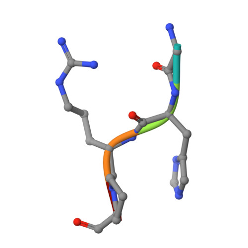
    Fibrinogen residue Bbeta432Asp is part of hole "b" that interacts with knob "B," whose sequence starts with Gly-His-Arg-Pro-amide (GHRP). Because previous studies showed BbetaD432A has normal polymerization, we hypothesized that Bbeta432Asp is not critical for knob "B" binding and that new knob-hole interactions would compensate for the loss of this Asp residue. To test this hypothesis, we solved the crystal structure of fragment D from BbetaD432A. Surprisingly, the structure (rfD-BbetaD432A+GH) showed the peptide GHRP was not bound to hole "b." We then re-evaluated the polymerization of this variant by examining clot turbidity, clot structure, and the rate of FXIIIa cross-linking. The turbidity and the rate of gamma-gamma dimer formation for BbetaD432A were indistinguishable compared with normal fibrinogen. Scanning electron microscopy showed no significant differences between the clots of BbetaD432A and normal, but the thrombin-derived clots had thicker fibers than clots obtained from batroxobin, suggesting that cleavage of FpB is more important than "B:b" interactions. We conclude that hole "b" and "B:b" knob-hole binding per se have no influence on fibrin polymerization.


  • Organizational Affiliation

    Department of Chemistry, University of North Carolina, Chapel Hill, NC 27599-7525, USA.


Macromolecules
Find similar proteins by:  (by identity cutoff)  |  3D Structure
Entity ID: 1
MoleculeChains Sequence LengthOrganismDetailsImage
Fibrinogen alpha chain
A, D
87Homo sapiensMutation(s): 0 
Gene Names: FGA
UniProt & NIH Common Fund Data Resources
Find proteins for P02671 (Homo sapiens)
Explore P02671 
Go to UniProtKB:  P02671
PHAROS:  P02671
GTEx:  ENSG00000171560 
Entity Groups  
Sequence Clusters30% Identity50% Identity70% Identity90% Identity95% Identity100% Identity
UniProt GroupP02671
Sequence Annotations
Expand
  • Reference Sequence
Find similar proteins by:  (by identity cutoff)  |  3D Structure
Entity ID: 2
MoleculeChains Sequence LengthOrganismDetailsImage
Fibrinogen beta chain
B, E
328Homo sapiensMutation(s): 1 
Gene Names: FGB
UniProt & NIH Common Fund Data Resources
Find proteins for P02675 (Homo sapiens)
Explore P02675 
Go to UniProtKB:  P02675
PHAROS:  P02675
GTEx:  ENSG00000171564 
Entity Groups  
Sequence Clusters30% Identity50% Identity70% Identity90% Identity95% Identity100% Identity
UniProt GroupP02675
Sequence Annotations
Expand
  • Reference Sequence
Find similar proteins by:  (by identity cutoff)  |  3D Structure
Entity ID: 3
MoleculeChains Sequence LengthOrganismDetailsImage
Fibrinogen gamma chain
C, F
319Homo sapiensMutation(s): 0 
Gene Names: FGGPRO2061
UniProt & NIH Common Fund Data Resources
Find proteins for P02679 (Homo sapiens)
Explore P02679 
Go to UniProtKB:  P02679
PHAROS:  P02679
GTEx:  ENSG00000171557 
Entity Groups  
Sequence Clusters30% Identity50% Identity70% Identity90% Identity95% Identity100% Identity
UniProt GroupP02679
Sequence Annotations
Expand
  • Reference Sequence

Find similar proteins by:  Sequence   |   3D Structure  

Entity ID: 4
MoleculeChains Sequence LengthOrganismDetailsImage
Gly-His-Arg-Pro-amide
G, H
5synthetic constructMutation(s): 0 
Sequence Annotations
Expand
  • Reference Sequence
Small Molecules
Ligands 2 Unique
IDChains Name / Formula / InChI Key2D Diagram3D Interactions
NAG
Query on NAG

Download Ideal Coordinates CCD File 
I [auth B],
N [auth E]
2-acetamido-2-deoxy-beta-D-glucopyranose
C8 H15 N O6
OVRNDRQMDRJTHS-FMDGEEDCSA-N
CA
Query on CA

Download Ideal Coordinates CCD File 
J [auth B]
K [auth B]
L [auth C]
M [auth C]
O [auth E]
J [auth B],
K [auth B],
L [auth C],
M [auth C],
O [auth E],
P [auth E],
Q [auth F]
CALCIUM ION
Ca
BHPQYMZQTOCNFJ-UHFFFAOYSA-N
Experimental Data & Validation

Experimental Data

  • Method: X-RAY DIFFRACTION
  • Resolution: 2.30 Å
  • R-Value Free: 0.242 
  • R-Value Work: 0.214 
  • R-Value Observed: 0.216 
  • Space Group: P 21 21 21
Unit Cell:
Length ( Å )Angle ( ˚ )
a = 54.476α = 90
b = 146.596β = 90
c = 228.96γ = 90
Software Package:
Software NamePurpose
REFMACrefinement
MOLREPphasing
CNSrefinement
HKL-2000data reduction
HKL-2000data scaling
HKL-2000data collection

Structure Validation

View Full Validation Report


Ligand Structure Quality Assessment 


Entry History 

Deposition Data

Revision History  (Full details and data files)

  • Version 1.0: 2009-01-13
    Type: Initial release
  • Version 1.1: 2011-07-13
    Changes: Advisory, Version format compliance
  • Version 1.2: 2013-09-18
    Changes: Derived calculations
  • Version 1.3: 2019-07-24
    Changes: Data collection, Refinement description, Source and taxonomy, Structure summary
  • Version 1.4: 2020-07-29
    Type: Remediation
    Reason: Carbohydrate remediation
    Changes: Advisory, Data collection, Derived calculations, Structure summary
  • Version 1.5: 2021-10-20
    Changes: Database references, Structure summary
  • Version 1.6: 2023-08-30
    Changes: Data collection, Refinement description