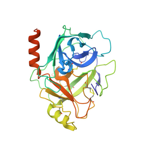
3E16

X-ray structure of human prostasin in complex with Benzoxazole warhead peptidomimic, lysine in P3


Experimental Data Snapshot

  • Method: X-RAY DIFFRACTION
  • Resolution: 1.60 Å
  • R-Value Free: 0.229 
  • R-Value Work: 0.185 
  • R-Value Observed: 0.188 

wwPDB Validation   3D Report Full Report


Ligand Structure Quality Assessment 


This is version 1.3 of the entry. See complete history


Literature

Discovery of inhibitors of the channel-activating protease prostasin (CAP1/PRSS8) utilizing structure-based design.

Tully, D.C.Vidal, A.Chatterjee, A.K.Williams, J.A.Roberts, M.J.Petrassi, H.M.Spraggon, G.Bursulaya, B.Pacoma, R.Shipway, A.Schumacher, A.M.Danahay, H.Harris, J.L.

(2008) Bioorg Med Chem Lett 18: 5895-5899

  • DOI: https://doi.org/10.1016/j.bmcl.2008.08.029
  • Primary Citation of Related Structures:  
    3E0P, 3E16

  • PubMed Abstract: 

    Structure-based design was utilized to guide the early stage optimization of a substrate-like inhibitor to afford potent peptidomimetic inhibitors of the channel-activating protease prostasin. The first X-ray crystal structures of prostasin with small molecule inhibitors bound to the active site are also reported.


  • Organizational Affiliation

    Genomics Institute of the Novartis Research Foundation, 10675 John J. Hopkins Dr., San Diego, CA 92121, USA. dtully@gnf.org


Macromolecules
Find similar proteins by:  (by identity cutoff)  |  3D Structure
Entity ID: 1
MoleculeChains Sequence LengthOrganismDetailsImage
ProstasinA [auth B]271Homo sapiensMutation(s): 3 
Gene Names: PRSS8
EC: 3.4.21
UniProt & NIH Common Fund Data Resources
Find proteins for Q16651 (Homo sapiens)
Explore Q16651 
Go to UniProtKB:  Q16651
PHAROS:  Q16651
GTEx:  ENSG00000052344 
Entity Groups  
Sequence Clusters30% Identity50% Identity70% Identity90% Identity95% Identity100% Identity
UniProt GroupQ16651
Sequence Annotations
Expand
  • Reference Sequence
Small Molecules
Ligands 3 Unique
IDChains Name / Formula / InChI Key2D Diagram3D Interactions
B4C
Query on B4C

Download Ideal Coordinates CCD File 
C [auth B]benzyl [(1S)-5-amino-1-{[(1S)-1-({(1S)-5-amino-1-[(S)-1,3-benzoxazol-2-yl(hydroxy)methyl]pentyl}carbamoyl)-3-phenylpropyl]carbamoyl}pentyl]carbamate
C37 H48 N6 O6
HJDPWVALYXRQIB-QUUJSONZSA-N
GOL
Query on GOL

Download Ideal Coordinates CCD File 
B
GLYCEROL
C3 H8 O3
PEDCQBHIVMGVHV-UHFFFAOYSA-N
DMS
Query on DMS

Download Ideal Coordinates CCD File 
D [auth B]DIMETHYL SULFOXIDE
C2 H6 O S
IAZDPXIOMUYVGZ-UHFFFAOYSA-N
Experimental Data & Validation

Experimental Data

  • Method: X-RAY DIFFRACTION
  • Resolution: 1.60 Å
  • R-Value Free: 0.229 
  • R-Value Work: 0.185 
  • R-Value Observed: 0.188 
  • Space Group: P 21 21 21
Unit Cell:
Length ( Å )Angle ( ˚ )
a = 53.253α = 90
b = 55.009β = 90
c = 82.799γ = 90
Software Package:
Software NamePurpose
PHENIXrefinement
ADSCdata collection
HKL-2000data reduction
HKL-2000data scaling

Structure Validation

View Full Validation Report


Ligand Structure Quality Assessment 


Entry History 

Deposition Data

Revision History  (Full details and data files)

  • Version 1.0: 2008-09-09
    Type: Initial release
  • Version 1.1: 2011-07-13
    Changes: Non-polymer description, Version format compliance
  • Version 1.2: 2021-07-28
    Changes: Database references, Derived calculations, Refinement description
  • Version 1.3: 2021-10-20
    Changes: Database references