3E0N

The X-ray structure of Human Prostasin in complex with DFFR-chloromethyl ketone inhibitor


Experimental Data Snapshot

  • Method: X-RAY DIFFRACTION
  • Resolution: 1.70 Å
  • R-Value Free: 0.202 
  • R-Value Work: 0.157 
  • R-Value Observed: 0.159 

wwPDB Validation   3D Report Full Report


This is version 2.1 of the entry. See complete history


Literature

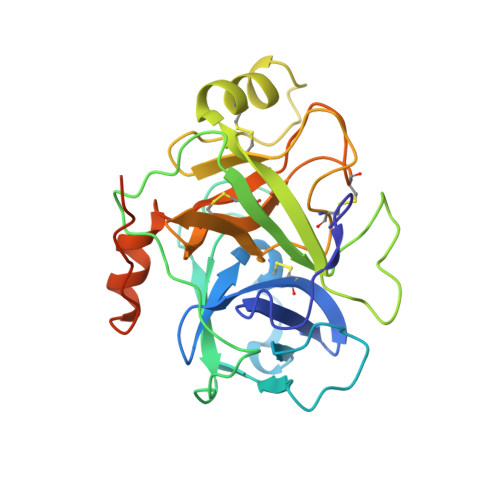
Active site conformational changes of prostasin provide a new mechanism of protease regulation by divalent cations.

Spraggon, G.Hornsby, M.Shipway, A.Tully, D.C.Bursulaya, B.Danahay, H.Harris, J.L.Lesley, S.A.

(2009) Protein Sci 18: 1081-1094

  • DOI: https://doi.org/10.1002/pro.118
  • Primary Citation of Related Structures:  
    3E0N, 3E1X, 3FVF, 3GYL, 3GYM

  • PubMed Abstract: 

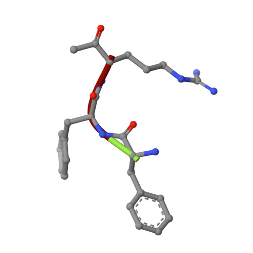
    Prostasin or human channel-activating protease 1 has been reported to play a critical role in the regulation of extracellular sodium ion transport via its activation of the epithelial cell sodium channel. Here, the structure of the extracellular portion of the membrane associated serine protease has been solved to high resolution in complex with a nonselective d-FFR chloromethyl ketone inhibitor, in an apo form, in a form where the apo crystal has been soaked with the covalent inhibitor camostat and in complex with the protein inhibitor aprotinin. It was also crystallized in the presence of the divalent cation Ca(+2). Comparison of the structures with each other and with other members of the trypsin-like serine protease family reveals unique structural features of prostasin and a large degree of conformational variation within specificity determining loops. Of particular interest is the S1 subsite loop which opens and closes in response to basic residues or divalent ions, directly binding Ca(+2) cations. This induced fit active site provides a new possible mode of regulation of trypsin-like proteases adapted in particular to extracellular regions with variable ionic concentrations such as the outer membrane layer of the epithelial cell.


  • Organizational Affiliation

    Genomics Institute of the Novartis Research Foundation, 10675 John Jay Hopkins Drive, San Diego, California 92121, USA. gspraggo@gnf.org


Macromolecules
Find similar proteins by:  (by identity cutoff)  |  3D Structure
Entity ID: 1
MoleculeChains Sequence LengthOrganismDetailsImage
Prostasin heavy chainA [auth B]271Homo sapiensMutation(s): 0 
Gene Names: PRSS8
UniProt & NIH Common Fund Data Resources
Find proteins for Q16651 (Homo sapiens)
Explore Q16651 
Go to UniProtKB:  Q16651
PHAROS:  Q16651
GTEx:  ENSG00000052344 
Entity Groups  
Sequence Clusters30% Identity50% Identity70% Identity90% Identity95% Identity100% Identity
UniProt GroupQ16651
Sequence Annotations
Expand
  • Reference Sequence

Find similar proteins by:  Sequence   |   3D Structure  

Entity ID: 2
MoleculeChains Sequence LengthOrganismDetailsImage
DPN-PHE-ARMB [auth A]3synthetic constructMutation(s): 0 
Sequence Annotations
Expand
  • Reference Sequence
Biologically Interesting Molecules (External Reference) 1 Unique
Experimental Data & Validation

Experimental Data

  • Method: X-RAY DIFFRACTION
  • Resolution: 1.70 Å
  • R-Value Free: 0.202 
  • R-Value Work: 0.157 
  • R-Value Observed: 0.159 
  • Space Group: P 21 21 21
Unit Cell:
Length ( Å )Angle ( ˚ )
a = 37.677α = 90
b = 75.88β = 90
c = 79.179γ = 90
Software Package:
Software NamePurpose
PHENIXrefinement
ADSCdata collection
MOSFLMdata reduction
SCALAdata scaling
MOLREPphasing

Structure Validation

View Full Validation Report


Entry History 

Deposition Data

Revision History  (Full details and data files)

  • Version 1.0: 2009-06-16
    Type: Initial release
  • Version 1.1: 2011-07-13
    Changes: Non-polymer description, Version format compliance
  • Version 1.2: 2021-10-20
    Changes: Database references, Derived calculations
  • Version 2.0: 2023-02-22
    Changes: Advisory, Atomic model, Data collection, Database references, Derived calculations, Polymer sequence, Source and taxonomy, Structure summary
  • Version 2.1: 2023-09-20
    Changes: Data collection, Refinement description