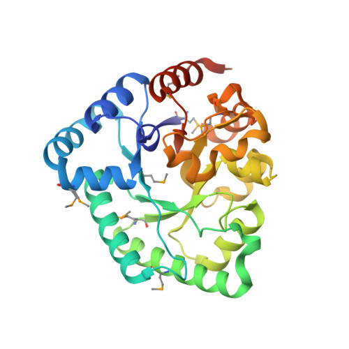
3DX5

Crystal structure of the probable 3-DHS dehydratase AsbF involved in the petrobactin synthesis from Bacillus anthracis


Experimental Data Snapshot

  • Method: X-RAY DIFFRACTION
  • Resolution: 2.12 Å
  • R-Value Free: 0.178 
  • R-Value Work: 0.148 
  • R-Value Observed: 0.150 

wwPDB Validation   3D Report Full Report


Ligand Structure Quality Assessment 


This is version 1.1 of the entry. See complete history


Literature

Structural and functional analysis of AsbF: origin of the stealth 3,4-dihydroxybenzoic acid subunit for petrobactin biosynthesis.

Pfleger, B.F.Kim, Y.Nusca, T.D.Maltseva, N.Lee, J.Y.Rath, C.M.Scaglione, J.B.Janes, B.K.Anderson, E.C.Bergman, N.H.Hanna, P.C.Joachimiak, A.Sherman, D.H.

(2008) Proc Natl Acad Sci U S A 105: 17133-17138

  • DOI: https://doi.org/10.1073/pnas.0808118105
  • Primary Citation of Related Structures:  
    3DX5

  • PubMed Abstract: 

    Petrobactin, a virulence-associated siderophore produced by Bacillus anthracis, chelates ferric iron through the rare 3,4-isomer of dihydroxybenzoic acid (3,4-DHBA). Most catechol siderophores, including bacillibactin and enterobactin, use 2,3-DHBA as a biosynthetic subunit. Significantly, siderocalin, a factor involved in human innate immunity, sequesters ferric siderophores bearing the more typical 2,3-DHBA moiety, thereby impeding uptake of iron by the pathogenic bacterial cell. In contrast, the unusual 3,4-DHBA component of petrobactin renders the siderocalin system incapable of obstructing bacterial iron uptake. Although recent genetic and biochemical studies have revealed selected early steps in petrobactin biosynthesis, the origin of 3,4-DHBA as well as the function of the protein encoded by the final gene in the B. anthracis siderophore biosynthetic (asb) operon, asbF (BA1986), has remained unclear. In this study we demonstrate that 3,4-DHBA is produced through conversion of the common bacterial metabolite 3-dehydroshikimate (3-DHS) by AsbF-a 3-DHS dehydratase. Elucidation of the cocrystal structure of AsbF with 3,4-DHBA, in conjunction with a series of biochemical studies, supports a mechanism in which an enolate intermediate is formed through the action of this 3-DHS dehydratase metalloenzyme. Structural and functional parallels are evident between AsbF and other enzymes within the xylose isomerase TIM-barrel family. Overall, these data indicate that microbial species shown to possess homologs of AsbF may, like B. anthracis, also rely on production of the unique 3,4-DHBA metabolite to achieve full viability in the environment or virulence within the host.


  • Organizational Affiliation

    Life Sciences Institute and Departments of Medicinal Chemistry and Chemistry, University of Michigan, 210 Washtenaw Avenue, Ann Arbor, MI 48109, USA.


Macromolecules
Find similar proteins by:  (by identity cutoff)  |  3D Structure
Entity ID: 1
MoleculeChains Sequence LengthOrganismDetailsImage
uncharacterized protein AsbF286Bacillus anthracisMutation(s): 0 
Gene Names: BAS1843BA_1986GBAA1986
UniProt
Find proteins for Q81RQ4 (Bacillus anthracis)
Explore Q81RQ4 
Go to UniProtKB:  Q81RQ4
Entity Groups  
Sequence Clusters30% Identity50% Identity70% Identity90% Identity95% Identity100% Identity
UniProt GroupQ81RQ4
Sequence Annotations
Expand
  • Reference Sequence
Small Molecules
Ligands 5 Unique
IDChains Name / Formula / InChI Key2D Diagram3D Interactions
DHB
Query on DHB

Download Ideal Coordinates CCD File 
D [auth A]3,4-DIHYDROXYBENZOIC ACID
C7 H6 O4
YQUVCSBJEUQKSH-UHFFFAOYSA-N
TRS
Query on TRS

Download Ideal Coordinates CCD File 
E [auth A]2-AMINO-2-HYDROXYMETHYL-PROPANE-1,3-DIOL
C4 H12 N O3
LENZDBCJOHFCAS-UHFFFAOYSA-O
GOL
Query on GOL

Download Ideal Coordinates CCD File 
F [auth A]GLYCEROL
C3 H8 O3
PEDCQBHIVMGVHV-UHFFFAOYSA-N
MN
Query on MN

Download Ideal Coordinates CCD File 
B [auth A]MANGANESE (II) ION
Mn
WAEMQWOKJMHJLA-UHFFFAOYSA-N
CL
Query on CL

Download Ideal Coordinates CCD File 
C [auth A]CHLORIDE ION
Cl
VEXZGXHMUGYJMC-UHFFFAOYSA-M
Modified Residues  1 Unique
IDChains TypeFormula2D DiagramParent
MSE
Query on MSE
A
L-PEPTIDE LINKINGC5 H11 N O2 SeMET
Experimental Data & Validation

Experimental Data

  • Method: X-RAY DIFFRACTION
  • Resolution: 2.12 Å
  • R-Value Free: 0.178 
  • R-Value Work: 0.148 
  • R-Value Observed: 0.150 
  • Space Group: P 65 2 2
Unit Cell:
Length ( Å )Angle ( ˚ )
a = 134.483α = 90
b = 134.483β = 90
c = 72.806γ = 120
Software Package:
Software NamePurpose
REFMACrefinement
SBC-Collectdata collection
HKL-3000data collection
HKL-3000data reduction
HKL-3000data scaling
HKL-3000phasing
MLPHAREphasing
DMphasing
SHELXDphasing
RESOLVEphasing
Cootmodel building

Structure Validation

View Full Validation Report


Ligand Structure Quality Assessment 


Entry History 

Revision History  (Full details and data files)

  • Version 1.0: 2008-09-02
    Type: Initial release
  • Version 1.1: 2011-07-13
    Changes: Advisory, Version format compliance