3DJL

Crystal structure of alkylation response protein E. coli AidB


Experimental Data Snapshot

  • Method: X-RAY DIFFRACTION
  • Resolution: 1.70 Å
  • R-Value Free: 0.179 
  • R-Value Work: 0.161 
  • R-Value Observed: 0.162 

wwPDB Validation   3D Report Full Report


Ligand Structure Quality Assessment 


This is version 1.2 of the entry. See complete history


Literature

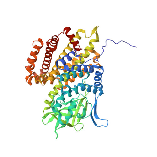
Structure and DNA binding of alkylation response protein AidB.

Bowles, T.Metz, A.H.O'Quin, J.Wawrzak, Z.Eichman, B.F.

(2008) Proc Natl Acad Sci U S A 105: 15299-15304

  • DOI: https://doi.org/10.1073/pnas.0806521105
  • Primary Citation of Related Structures:  
    3DJL

  • PubMed Abstract: 

    Exposure of Escherichia coli to alkylating agents activates expression of AidB in addition to DNA repair proteins Ada, AlkA, and AlkB. AidB was recently shown to possess a flavin adenine dinucleotide (FAD) cofactor and to bind to dsDNA, implicating it as a flavin-dependent DNA repair enzyme. However, the molecular mechanism by which AidB acts to reduce the mutagenic effects of specific DNA alkylators is unknown. We present a 1.7-A crystal structure of AidB, which bears superficial resemblance to the acyl-CoA dehydrogenase superfamily of flavoproteins. The structure reveals a unique quaternary organization and a distinctive FAD active site that provides a rationale for AidB's limited dehydrogenase activity. A highly electropositive C-terminal domain not present in structural homologs was identified by mutational analysis as the DNA binding site. Structural analysis of the DNA and FAD binding sites provides evidence against AidB-catalyzed DNA repair and supports a model in which AidB acts to prevent alkylation damage by protecting DNA and destroying alkylating agents that have yet to reach their DNA target.


  • Organizational Affiliation

    Department of Biological Sciences and Center for Structural Biology, Vanderbilt University, Nashville, TN 37232, USA.


Macromolecules
Find similar proteins by:  (by identity cutoff)  |  3D Structure
Entity ID: 1
MoleculeChains Sequence LengthOrganismDetailsImage
Protein aidB541Escherichia coliMutation(s): 0 
Gene Names: aidB
UniProt
Find proteins for P33224 (Escherichia coli (strain K12))
Explore P33224 
Go to UniProtKB:  P33224
Entity Groups  
Sequence Clusters30% Identity50% Identity70% Identity90% Identity95% Identity100% Identity
UniProt GroupP33224
Sequence Annotations
Expand
  • Reference Sequence
Small Molecules
Ligands 2 Unique
IDChains Name / Formula / InChI Key2D Diagram3D Interactions
FAD
Query on FAD

Download Ideal Coordinates CCD File 
C [auth A]FLAVIN-ADENINE DINUCLEOTIDE
C27 H33 N9 O15 P2
VWWQXMAJTJZDQX-UYBVJOGSSA-N
CA
Query on CA

Download Ideal Coordinates CCD File 
B [auth A]CALCIUM ION
Ca
BHPQYMZQTOCNFJ-UHFFFAOYSA-N
Experimental Data & Validation

Experimental Data

  • Method: X-RAY DIFFRACTION
  • Resolution: 1.70 Å
  • R-Value Free: 0.179 
  • R-Value Work: 0.161 
  • R-Value Observed: 0.162 
  • Space Group: I 2 2 2
Unit Cell:
Length ( Å )Angle ( ˚ )
a = 84.989α = 90
b = 101.875β = 90
c = 137.566γ = 90
Software Package:
Software NamePurpose
REFMACrefinement
SERGUIdata collection
HKL-2000data reduction
HKL-2000data scaling
SHARPphasing

Structure Validation

View Full Validation Report


Ligand Structure Quality Assessment 


Entry History 

Deposition Data

Revision History  (Full details and data files)

  • Version 1.0: 2008-09-23
    Type: Initial release
  • Version 1.1: 2011-07-13
    Changes: Advisory, Version format compliance
  • Version 1.2: 2012-03-21
    Changes: Database references, Derived calculations