3D9A

High Resolution Crystal Structure Structure of HyHel10 Fab Complexed to Hen Egg Lysozyme


Experimental Data Snapshot

  • Method: X-RAY DIFFRACTION
  • Resolution: 1.20 Å
  • R-Value Free: 0.205 
  • R-Value Work: 0.190 
  • R-Value Observed: 0.191 

wwPDB Validation   3D Report Full Report


This is version 1.4 of the entry. See complete history


Literature

Light chain somatic mutations change thermodynamics of binding and water coordination in the HyHEL-10 family of antibodies.

Acchione, M.Lipschultz, C.A.DeSantis, M.E.Shanmuganathan, A.Li, M.Wlodawer, A.Tarasov, S.Smith-Gill, S.J.

(2009) Mol Immunol 47: 457-464

  • DOI: https://doi.org/10.1016/j.molimm.2009.08.018
  • Primary Citation of Related Structures:  
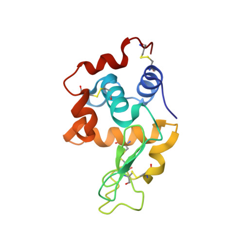
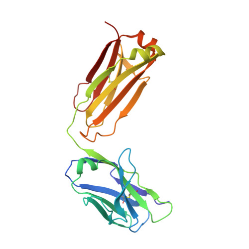
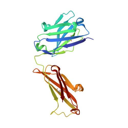
    3D9A

  • PubMed Abstract: 

    Thermodynamic and structural studies addressed the increased affinity due to L-chain somatic mutations in the HyHEL-10 family of affinity matured IgG antibodies, using ITC, SPR with van't Hoff analysis, and X-ray crystallography. When compared to the parental antibody H26L26, the H26L10 and H26L8 chimeras binding to lysozyme showed an increase in favorable DeltaG(o) of -1.2+/-0.1 kcal mol(-1) and -1.3+/-0.1 kcal mol(-1), respectively. Increase in affinity of the H26L10 chimera was due to a net increase in favorable enthalpy change with little difference in change in entropy compared to H26L26. The H26L8 chimera exhibited the greatest increase in favorable enthalpy but also showed an increase in unfavorable entropy change, with the result being that the affinities of both chimeras were essentially equivalent. Site-directed L-chain mutants identified the shared somatic mutation S30G as the dominant contributor to increasing affinity to lysozyme. This mutation was not influenced by H-chain somatic mutations. Residue 30L is at the periphery of the binding interface and S30G effects an increase in hydrophobicity and decrease in H-bonding ability and size, but does not make any new energetically important antigen contacts. A new 1.2-A structure of the H10L10-HEL complex showed changes in the pattern of both inter- and intra-molecular water bridging with no other significant structural alterations near the binding interface compared to the H26L26-HEL complex. These results highlight the necessity for investigating both the structure and the thermodynamics associated with introduced mutations, in order to better assess and understand their impact on binding. Furthermore, it provides an important example of how backbone flexibility and water-bridging may favorably influence the thermodynamics of an antibody-antigen interaction.


  • Organizational Affiliation

    Structural Biophysics Laboratory, Center for Cancer Research, National Cancer Institute, Frederick, MD, USA.


Macromolecules
Find similar proteins by:  (by identity cutoff)  |  3D Structure
Entity ID: 1
MoleculeChains Sequence LengthOrganismDetailsImage
Lysozyme CA [auth C]129Gallus gallusMutation(s): 0 
EC: 3.2.1.17
UniProt
Find proteins for P00698 (Gallus gallus)
Explore P00698 
Go to UniProtKB:  P00698
Entity Groups  
Sequence Clusters30% Identity50% Identity70% Identity90% Identity95% Identity100% Identity
UniProt GroupP00698
Sequence Annotations
Expand
  • Reference Sequence
Find similar proteins by:  (by identity cutoff)  |  3D Structure
Entity ID: 2
MoleculeChains Sequence LengthOrganismDetailsImage
Light Chain of HyHel10 Antibody Fragment (Fab)B [auth L]213Mus musculusMutation(s): 0 
Entity Groups  
Sequence Clusters30% Identity50% Identity70% Identity90% Identity95% Identity100% Identity
Sequence Annotations
Expand
  • Reference Sequence
Find similar proteins by:  (by identity cutoff)  |  3D Structure
Entity ID: 3
MoleculeChains Sequence LengthOrganismDetailsImage
Heavy Chain of HyHel10 Antibody Fragment (Fab)C [auth H]210Mus musculusMutation(s): 0 
Entity Groups  
Sequence Clusters30% Identity50% Identity70% Identity90% Identity95% Identity100% Identity
Sequence Annotations
Expand
  • Reference Sequence
Experimental Data & Validation

Experimental Data

  • Method: X-RAY DIFFRACTION
  • Resolution: 1.20 Å
  • R-Value Free: 0.205 
  • R-Value Work: 0.190 
  • R-Value Observed: 0.191 
  • Space Group: P 1 21 1
Unit Cell:
Length ( Å )Angle ( ˚ )
a = 39.842α = 90
b = 77.385β = 96.66
c = 89.115γ = 90
Software Package:
Software NamePurpose
DENZOdata reduction
SCALEPACKdata scaling
PHASERphasing
REFMACrefinement
PDB_EXTRACTdata extraction

Structure Validation

View Full Validation Report


Entry History 

Deposition Data

Revision History  (Full details and data files)

  • Version 1.0: 2008-06-10
    Type: Initial release
  • Version 1.1: 2011-07-13
    Changes: Advisory, Version format compliance
  • Version 1.2: 2013-09-25
    Changes: Derived calculations
  • Version 1.3: 2017-10-25
    Changes: Advisory, Refinement description
  • Version 1.4: 2023-08-30
    Changes: Data collection, Database references, Refinement description