3D24

Crystal structure of ligand-binding domain of estrogen-related receptor alpha (ERRalpha) in complex with the peroxisome proliferators-activated receptor coactivator-1alpha box3 peptide (PGC-1alpha)


Experimental Data Snapshot

  • Method: X-RAY DIFFRACTION
  • Resolution: 2.11 Å
  • R-Value Free: 0.255 
  • R-Value Work: 0.212 
  • R-Value Observed: 0.212 

wwPDB Validation   3D Report Full Report


This is version 1.2 of the entry. See complete history


Literature

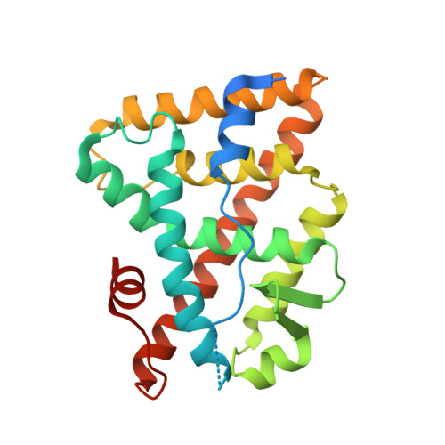
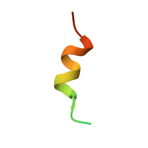
Communication between the ERR{alpha} Homodimer Interface and the PGC-1{alpha} Binding Surface via the Helix 8-9 Loop.

Greschik, H.Althage, M.Flaig, R.Sato, Y.Chavant, V.Peluso-Iltis, C.Choulier, L.Cronet, P.Rochel, N.Schule, R.Stromstedt, P.E.Moras, D.

(2008) J Biol Chem 283: 20220-20230

  • DOI: https://doi.org/10.1074/jbc.M801920200
  • Primary Citation of Related Structures:  
    3D24

  • PubMed Abstract: 

    Although structural studies on the ligand-binding domain (LBD) have established the general mode of nuclear receptor (NR)/coactivator interaction, determinants of binding specificity are only partially understood. The LBD of estrogen receptor-alpha (ERalpha), for example, interacts only with a region of peroxisome proliferator-activated receptor coactivator (PGC)-1alpha, which contains the canonical LXXLL motif (NR box2), whereas the LBD of estrogen-related receptor-alpha (ERRalpha) also binds efficiently an untypical, LXXYL-containing region (NR box3) of PGC-1alpha. Surprisingly, in a previous structural study, the ERalpha LBD has been observed to bind NR box3 of transcriptional intermediary factor (TIF)-2 untypically via LXXYL, whereas the ERRalpha LBD binds this region of TIF-2 only poorly. Here we present a new crystal structure of the ERRalpha LBD in complex with a PGC-1alpha box3 peptide. In this structure, residues N-terminal of the PGC-1alpha LXXYL motif formed contacts with helix 4, the loop connecting helices 8 and 9, and with the C terminus of the ERRalpha LBD. Interaction studies using wild-type and mutant PGC-1alpha and ERRalpha showed that these contacts are functionally relevant and are required for efficient ERRalpha/PGC-1alpha interaction. Furthermore, a structure comparison between ERRalpha and ERalpha and mutation analyses provided evidence that the helix 8-9 loop, which differs significantly in both nuclear receptors, is a major determinant of coactivator binding specificity. Finally, our results revealed that in ERRalpha the helix 8-9 loop allosterically links the LBD homodimer interface with the coactivator cleft, thus providing a plausible explanation for distinct PGC-1alpha binding to ERRalpha monomers and homodimers.


  • Organizational Affiliation

    Département de Biologie et Génomique Structurales, Institut de Génétique et de Biologie Moléculaire et Cellulaire, Illkirch, France.


Macromolecules
Find similar proteins by:  (by identity cutoff)  |  3D Structure
Entity ID: 1
MoleculeChains Sequence LengthOrganismDetailsImage
Steroid hormone receptor ERR1
A, C
253Homo sapiensMutation(s): 0 
Gene Names: ESRRAERR1ESRL1NR3B1
UniProt & NIH Common Fund Data Resources
Find proteins for P11474 (Homo sapiens)
Explore P11474 
Go to UniProtKB:  P11474
PHAROS:  P11474
GTEx:  ENSG00000173153 
Entity Groups  
Sequence Clusters30% Identity50% Identity70% Identity90% Identity95% Identity100% Identity
UniProt GroupP11474
Sequence Annotations
Expand
  • Reference Sequence

Find similar proteins by:  Sequence   |   3D Structure  

Entity ID: 2
MoleculeChains Sequence LengthOrganismDetailsImage
Peroxisome proliferator-activated receptor gamma coactivator 1-alpha
B, D
22N/AMutation(s): 0 
UniProt & NIH Common Fund Data Resources
Find proteins for Q9UBK2 (Homo sapiens)
Explore Q9UBK2 
Go to UniProtKB:  Q9UBK2
PHAROS:  Q9UBK2
GTEx:  ENSG00000109819 
Entity Groups  
Sequence Clusters30% Identity50% Identity70% Identity90% Identity95% Identity100% Identity
UniProt GroupQ9UBK2
Sequence Annotations
Expand
  • Reference Sequence
Experimental Data & Validation

Experimental Data

  • Method: X-RAY DIFFRACTION
  • Resolution: 2.11 Å
  • R-Value Free: 0.255 
  • R-Value Work: 0.212 
  • R-Value Observed: 0.212 
  • Space Group: C 1 2 1
Unit Cell:
Length ( Å )Angle ( ˚ )
a = 119.4α = 90
b = 56β = 106.4
c = 96.2γ = 90
Software Package:
Software NamePurpose
HKL-2000data collection
AMoREphasing
CNSrefinement
HKL-2000data reduction
HKL-2000data scaling

Structure Validation

View Full Validation Report


Entry History 

Deposition Data

Revision History  (Full details and data files)

  • Version 1.0: 2008-06-10
    Type: Initial release
  • Version 1.1: 2011-07-13
    Changes: Version format compliance
  • Version 1.2: 2023-08-30
    Changes: Data collection, Database references, Refinement description