3CZP

Crystal structure of putative polyphosphate kinase 2 from Pseudomonas aeruginosa PA01


Experimental Data Snapshot

  • Method: X-RAY DIFFRACTION
  • Resolution: 2.00 Å
  • R-Value Free: 0.231 
  • R-Value Work: 0.173 
  • R-Value Observed: 0.176 

wwPDB Validation   3D Report Full Report


This is version 1.1 of the entry. See complete history


Literature

Polyphosphate-dependent synthesis of ATP and ADP by the family-2 polyphosphate kinases in bacteria.

Nocek, B.Kochinyan, S.Proudfoot, M.Brown, G.Evdokimova, E.Osipiuk, J.Edwards, A.M.Savchenko, A.Joachimiak, A.Yakunin, A.F.

(2008) Proc Natl Acad Sci U S A 105: 17730-17735

  • DOI: https://doi.org/10.1073/pnas.0807563105
  • Primary Citation of Related Structures:  
    3CZP, 3CZQ

  • PubMed Abstract: 

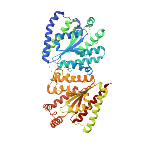
    Inorganic polyphosphate (polyP) is a linear polymer of tens or hundreds of phosphate residues linked by high-energy bonds. It is found in all organisms and has been proposed to serve as an energy source in a pre-ATP world. This ubiquitous and abundant biopolymer plays numerous and vital roles in metabolism and regulation in prokaryotes and eukaryotes, but the underlying molecular mechanisms for most activities of polyP remain unknown. In prokaryotes, the synthesis and utilization of polyP are catalyzed by 2 families of polyP kinases, PPK1 and PPK2, and polyphosphatases. Here, we present structural and functional characterization of the PPK2 family. Proteins with a single PPK2 domain catalyze polyP-dependent phosphorylation of ADP to ATP, whereas proteins containing 2 fused PPK2 domains phosphorylate AMP to ADP. Crystal structures of 2 representative proteins, SMc02148 from Sinorhizobium meliloti and PA3455 from Pseudomonas aeruginosa, revealed a 3-layer alpha/beta/alpha sandwich fold with an alpha-helical lid similar to the structures of microbial thymidylate kinases, suggesting that these proteins share a common evolutionary origin and catalytic mechanism. Alanine replacement mutagenesis identified 9 conserved residues, which are required for activity and include the residues from both Walker A and B motifs and the lid. Thus, the PPK2s represent a molecular mechanism, which potentially allow bacteria to use polyP as an intracellular energy reserve for the generation of ATP and survival.


  • Organizational Affiliation

    Midwest Center for Structural Genomics, Department of Biosciences, Argonne National Laboratory, 9700 South Cass Avenue, Building 202, Argonne, IL 60439, USA.


Macromolecules
Find similar proteins by:  (by identity cutoff)  |  3D Structure
Entity ID: 1
MoleculeChains Sequence LengthOrganismDetailsImage
Putative polyphosphate kinase 2
A, B
500Pseudomonas aeruginosa PAO1Mutation(s): 0 
Gene Names: PA3455
UniProt
Find proteins for Q9HYF1 (Pseudomonas aeruginosa (strain ATCC 15692 / DSM 22644 / CIP 104116 / JCM 14847 / LMG 12228 / 1C / PRS 101 / PAO1))
Explore Q9HYF1 
Go to UniProtKB:  Q9HYF1
Entity Groups  
Sequence Clusters30% Identity50% Identity70% Identity90% Identity95% Identity100% Identity
UniProt GroupQ9HYF1
Sequence Annotations
Expand
  • Reference Sequence
Small Molecules
Ligands 4 Unique
IDChains Name / Formula / InChI Key2D Diagram3D Interactions
MLI
Query on MLI

Download Ideal Coordinates CCD File 
M [auth B]MALONATE ION
C3 H2 O4
OFOBLEOULBTSOW-UHFFFAOYSA-L
GOL
Query on GOL

Download Ideal Coordinates CCD File 
H [auth A],
I [auth A],
J [auth A],
K [auth A]
GLYCEROL
C3 H8 O3
PEDCQBHIVMGVHV-UHFFFAOYSA-N
EDO
Query on EDO

Download Ideal Coordinates CCD File 
G [auth A],
N [auth B],
O [auth B]
1,2-ETHANEDIOL
C2 H6 O2
LYCAIKOWRPUZTN-UHFFFAOYSA-N
ACT
Query on ACT

Download Ideal Coordinates CCD File 
C [auth A],
D [auth A],
E [auth A],
F [auth A],
L [auth B]
ACETATE ION
C2 H3 O2
QTBSBXVTEAMEQO-UHFFFAOYSA-M
Modified Residues  1 Unique
IDChains TypeFormula2D DiagramParent
MSE
Query on MSE
A, B
L-PEPTIDE LINKINGC5 H11 N O2 SeMET
Experimental Data & Validation

Experimental Data

  • Method: X-RAY DIFFRACTION
  • Resolution: 2.00 Å
  • R-Value Free: 0.231 
  • R-Value Work: 0.173 
  • R-Value Observed: 0.176 
  • Space Group: P 21 21 21
Unit Cell:
Length ( Å )Angle ( ˚ )
a = 97.089α = 90
b = 100.735β = 90
c = 120.128γ = 90
Software Package:
Software NamePurpose
REFMACrefinement
SBC-Collectdata collection
HKL-3000data reduction
HKL-3000data scaling
HKL-3000phasing
SHELXDphasing
ARP/wARPmodel building
Cootmodel building
MLPHAREphasing
CCP4phasing

Structure Validation

View Full Validation Report


Entry History 

Revision History  (Full details and data files)

  • Version 1.0: 2008-07-01
    Type: Initial release
  • Version 1.1: 2011-07-13
    Changes: Advisory, Version format compliance