3CVI

How TCR-like antibody recognizes MHC-bound peptide


Experimental Data Snapshot

  • Method: X-RAY DIFFRACTION
  • Resolution: 1.80 Å
  • R-Value Free: 0.264 
  • R-Value Work: 0.219 

wwPDB Validation   3D Report Full Report


This is version 1.2 of the entry. See complete history


Literature

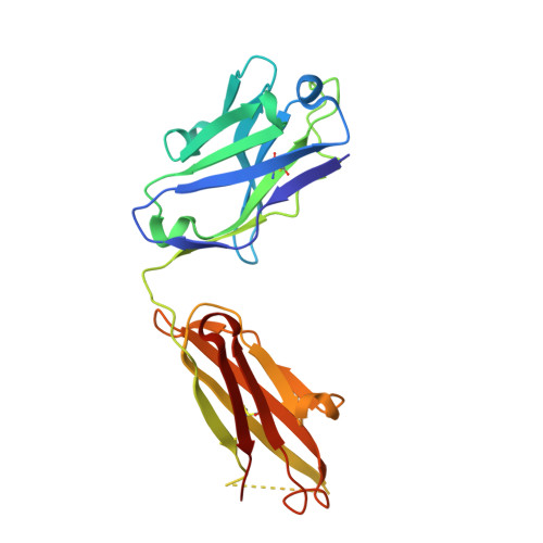
How a T cell receptor-like antibody recognizes major histocompatibility complex-bound peptide

Mareeva, T.Martinez-Hackert, E.Sykulev, Y.

(2008) J Biol Chem 283: 29053-29059

  • DOI: https://doi.org/10.1074/jbc.M804996200
  • Primary Citation of Related Structures:  
    3CVH, 3CVI

  • PubMed Abstract: 

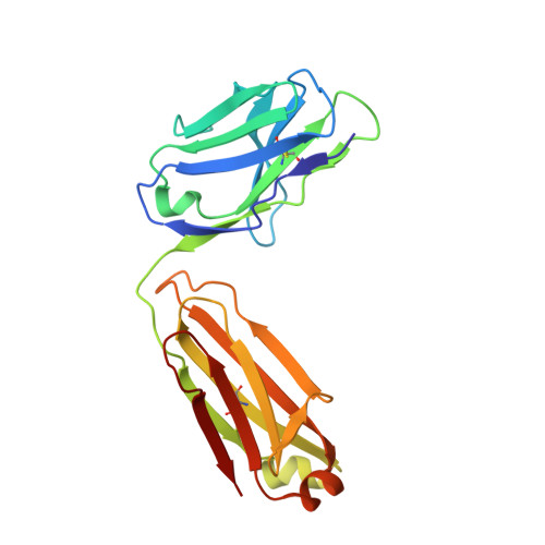
    We determined the crystal structures of the T cell receptor (TCR)-like antibody 25-D1.16 Fab fragment bound to a complex of SIINFEKL peptide from ovalbumin and the H-2K(b) molecule. Remarkably, this antibody directly "reads" the structure of the major histocompatibility complex (MHC)-bound peptide, employing the canonical diagonal binding mode utilized by most TCRs. This is in marked contrast with another TCR-like antibody, Hyb3, bound to melanoma peptide MAGE-A1 in association with HLA-A1 MHC class I. Hyb3 assumes a non-canonical orientation over its cognate peptide-MHC and appears to recognize a conformational epitope in which the MHC contribution is dominant. We conclude that TCR-like antibodies can recognize MHC-bound peptide via two different mechanisms: one is similar to that exploited by the preponderance of TCRs and the other requires a non-canonical antibody orientation over the peptide-MHC complex.


  • Organizational Affiliation

    Department of Microbiology and Immunology and the Kimmel Cancer Center, Thomas Jefferson University, Philadelphia, Pennsylvania 19107, USA.


Macromolecules
Find similar proteins by:  (by identity cutoff)  |  3D Structure
Entity ID: 1
MoleculeChains Sequence LengthOrganismDetailsImage
25-D1.16 Heavy chainA [auth H]219Mus musculusMutation(s): 0 
Entity Groups  
Sequence Clusters30% Identity50% Identity70% Identity90% Identity95% Identity100% Identity
Sequence Annotations
Expand
  • Reference Sequence
Find similar proteins by:  (by identity cutoff)  |  3D Structure
Entity ID: 2
MoleculeChains Sequence LengthOrganismDetailsImage
25-D1.16 Light chainB [auth L]209Mus musculusMutation(s): 0 
Entity Groups  
Sequence Clusters30% Identity50% Identity70% Identity90% Identity95% Identity100% Identity
Sequence Annotations
Expand
  • Reference Sequence
Experimental Data & Validation

Experimental Data

  • Method: X-RAY DIFFRACTION
  • Resolution: 1.80 Å
  • R-Value Free: 0.264 
  • R-Value Work: 0.219 
  • Space Group: P 1 21 1
Unit Cell:
Length ( Å )Angle ( ˚ )
a = 49.44α = 90
b = 60.86β = 111.97
c = 79.75γ = 90
Software Package:
Software NamePurpose
PHASERphasing
d*TREKdata scaling
CNSrefinement
PDB_EXTRACTdata extraction
CrystalCleardata collection
d*TREKdata reduction

Structure Validation

View Full Validation Report


Entry History 

Deposition Data

Revision History  (Full details and data files)

  • Version 1.0: 2008-09-23
    Type: Initial release
  • Version 1.1: 2011-07-13
    Changes: Version format compliance
  • Version 1.2: 2023-08-30
    Changes: Data collection, Database references, Refinement description