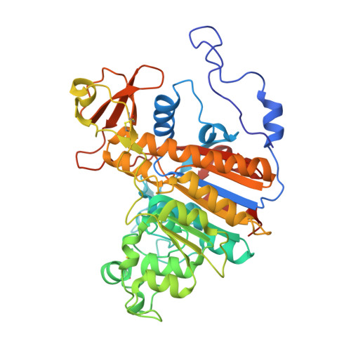
Arginine coordination in enzymatic phosphoryl transfer: evaluation of the effect of Arg166 mutations in Escherichia coli alkaline phosphatase
O'Brien, P.J., Lassila, J.K., Fenn, T.D., Zalatan, J.G., Herschlag, D.(2008) Biochemistry 47: 7663-7672
- PubMed: 18627128
- DOI: https://doi.org/10.1021/bi800545n
- Primary Citation of Related Structures:
3CMR - PubMed Abstract:
Arginine residues are commonly found in the active sites of enzymes catalyzing phosphoryl transfer reactions. Numerous site-directed mutagenesis experiments establish the importance of these residues for efficient catalysis, but their role in catalysis is not clear. To examine the role of arginine residues in the phosphoryl transfer reaction, we have measured the consequences of mutations to arginine 166 in Escherichia coli alkaline phosphatase on hydrolysis of ethyl phosphate, on individual reaction steps in the hydrolysis of the covalent enzyme-phosphoryl intermediate, and on thio substitution effects. The results show that the role of the arginine side chain extends beyond its positive charge, as the Arg166Lys mutant is as compromised in activity as Arg166Ser. Through measurement of individual reaction steps, we construct a free energy profile for the hydrolysis of the enzyme-phosphate intermediate. This analysis indicates that the arginine side chain strengthens binding by approximately 3 kcal/mol and provides an additional 1-2 kcal/mol stabilization of the chemical transition state. A 2.1 A X-ray diffraction structure of Arg166Ser AP is presented, which shows little difference in enzyme structure compared to the wild-type enzyme but shows a significant reorientation of the bound phosphate. Altogether, these results support a model in which the arginine contributes to catalysis through binding interactions and through additional transition state stabilization that may arise from complementarity of the guanidinum group to the geometry of the trigonal bipyramidal transition state.
Organizational Affiliation:
Department of Biochemistry, Stanford University, Stanford, California 94305, USA.