3CFD

Purple-fluorescent antibody EP2-25C10 in complex with its stilbene hapten


Experimental Data Snapshot

  • Method: X-RAY DIFFRACTION
  • Resolution: 2.50 Å
  • R-Value Free: 0.243 
  • R-Value Work: 0.211 
  • R-Value Observed: 0.212 

wwPDB Validation   3D Report Full Report


Ligand Structure Quality Assessment 


This is version 1.2 of the entry. See complete history


Literature

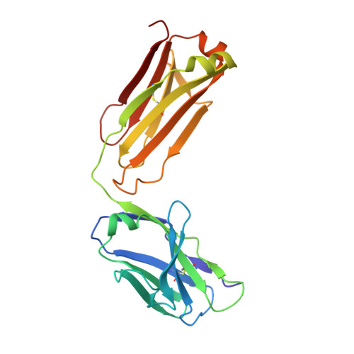
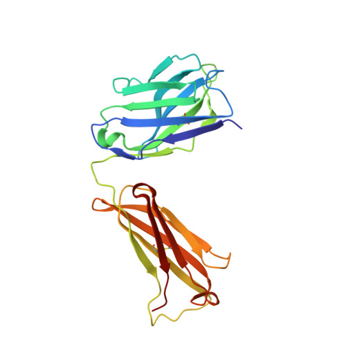
Deeply inverted electron-hole recombination in a luminescent antibody-stilbene complex.

Debler, E.W.Kaufmann, G.F.Meijler, M.M.Heine, A.Mee, J.M.Pljevaljcic, G.Di Bilio, A.J.Schultz, P.G.Millar, D.P.Janda, K.D.Wilson, I.A.Gray, H.B.Lerner, R.A.

(2008) Science 319: 1232-1235

  • DOI: https://doi.org/10.1126/science.1153445
  • Primary Citation of Related Structures:  
    3CFB, 3CFC, 3CFD, 3CFE

  • PubMed Abstract: 

    The blue-emissive antibody EP2-19G2 that has been elicited against trans-stilbene has unprecedented ability to produce bright luminescence and has been used as a biosensor in various applications. We show that the prolonged luminescence is not stilbene fluorescence. Instead, the emissive species is a charge-transfer excited complex of an anionic stilbene and a cationic, parallel pi-stacked tryptophan. Upon charge recombination, this complex generates exceptionally bright blue light. Complex formation is enabled by a deeply penetrating ligand-binding pocket, which in turn results from a noncanonical interface between the two variable domains of the antibody.


  • Organizational Affiliation

    Department of Molecular Biology, Scripps Research Institute, La Jolla, CA 92037, USA.


Macromolecules
Find similar proteins by:  (by identity cutoff)  |  3D Structure
Entity ID: 1
MoleculeChains Sequence LengthOrganismDetailsImage
PURPLE-FLUORESCENT ANTIBODY EP2-25C10-KAPPA LIGHT CHAINA [auth L],
C [auth A]
214Mus musculusMutation(s): 0 
Entity Groups  
Sequence Clusters30% Identity50% Identity70% Identity90% Identity95% Identity100% Identity
Sequence Annotations
Expand
  • Reference Sequence
Find similar proteins by:  (by identity cutoff)  |  3D Structure
Entity ID: 2
MoleculeChains Sequence LengthOrganismDetailsImage
PURPLE-FLUORESCENT ANTIBODY EP2-25C10-IGG2B HEAVY CHAINB [auth H],
D [auth B]
220Mus musculusMutation(s): 0 
Entity Groups  
Sequence Clusters30% Identity50% Identity70% Identity90% Identity95% Identity100% Identity
Sequence Annotations
Expand
  • Reference Sequence
Experimental Data & Validation

Experimental Data

  • Method: X-RAY DIFFRACTION
  • Resolution: 2.50 Å
  • R-Value Free: 0.243 
  • R-Value Work: 0.211 
  • R-Value Observed: 0.212 
  • Space Group: P 1
Unit Cell:
Length ( Å )Angle ( ˚ )
a = 59.99α = 91.98
b = 72.48β = 106.11
c = 77.23γ = 114.15
Software Package:
Software NamePurpose
PHASERphasing
REFMACrefinement
HKL-2000data reduction
HKL-2000data scaling

Structure Validation

View Full Validation Report


Ligand Structure Quality Assessment 


Entry History 

Deposition Data

Revision History  (Full details and data files)

  • Version 1.0: 2008-03-18
    Type: Initial release
  • Version 1.1: 2011-07-13
    Changes: Advisory, Version format compliance
  • Version 1.2: 2023-08-30
    Changes: Data collection, Database references, Derived calculations, Refinement description