3CF6

Structure of Epac2 in complex with cyclic-AMP and Rap


Experimental Data Snapshot

  • Method: X-RAY DIFFRACTION
  • Resolution: 2.20 Å
  • R-Value Free: 0.265 
  • R-Value Work: 0.243 
  • R-Value Observed: 0.244 

wwPDB Validation   3D Report Full Report


Ligand Structure Quality Assessment 


This is version 1.4 of the entry. See complete history


Literature

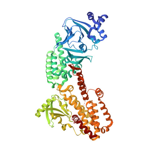
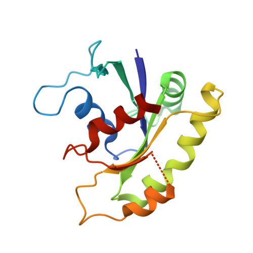
Structure of Epac2 in complex with a cyclic AMP analogue and RAP1B

Rehmann, H.Arias-Palomo, E.Hadders, M.A.Schwede, F.Llorca, O.Bos, J.L.

(2008) Nature 455: 124-127

  • DOI: https://doi.org/10.1038/nature07187
  • Primary Citation of Related Structures:  
    3CF6

  • PubMed Abstract: 

    Epac proteins are activated by binding of the second messenger cAMP and then act as guanine nucleotide exchange factors for Rap proteins. The Epac proteins are involved in the regulation of cell adhesion and insulin secretion. Here we have determined the structure of Epac2 in complex with a cAMP analogue (Sp-cAMPS) and RAP1B by X-ray crystallography and single particle electron microscopy. The structure represents the cAMP activated state of the Epac2 protein with the RAP1B protein trapped in the course of the exchange reaction. Comparison with the inactive conformation reveals that cAMP binding causes conformational changes that allow the cyclic nucleotide binding domain to swing from a position blocking the Rap binding site towards a docking site at the Ras exchange motif domain.


  • Organizational Affiliation

    Department of Physiological Chemistry, Centre for Biomedical Genetics and Cancer Genomics Centre, University Medical Center, Universiteitsweg 100, 3584 CG Utrecht, The Netherlands. h.rehmann@UMCutrecht.nl


Macromolecules
Find similar proteins by:  (by identity cutoff)  |  3D Structure
Entity ID: 1
MoleculeChains Sequence LengthOrganismDetailsImage
Rap guanine nucleotide exchange factor (GEF) 4A [auth E]694Mus musculusMutation(s): 0 
Gene Names: Rapgef4
Entity Groups  
Sequence Clusters30% Identity50% Identity70% Identity90% Identity95% Identity100% Identity
Sequence Annotations
Expand
  • Reference Sequence
Find similar proteins by:  (by identity cutoff)  |  3D Structure
Entity ID: 2
MoleculeChains Sequence LengthOrganismDetailsImage
Ras-related protein Rap-1bB [auth R]167Homo sapiensMutation(s): 0 
Gene Names: RAP1B
UniProt & NIH Common Fund Data Resources
Find proteins for P61224 (Homo sapiens)
Explore P61224 
Go to UniProtKB:  P61224
PHAROS:  P61224
GTEx:  ENSG00000127314 
Entity Groups  
Sequence Clusters30% Identity50% Identity70% Identity90% Identity95% Identity100% Identity
UniProt GroupP61224
Sequence Annotations
Expand
  • Reference Sequence
Small Molecules
Ligands 2 Unique
IDChains Name / Formula / InChI Key2D Diagram3D Interactions
SP1
Query on SP1

Download Ideal Coordinates CCD File 
C [auth E],
D [auth E]
6-(6-AMINO-PURIN-9-YL)-2-THIOXO-TETRAHYDRO-2-FURO[3,2-D][1,3,2]DIOXAPHOSPHININE-2,7-DIOL
C10 H12 N5 O5 P S
SMPNJFHAPJOHPP-LHKKBNDGSA-N
SO4
Query on SO4

Download Ideal Coordinates CCD File 
E [auth R]SULFATE ION
O4 S
QAOWNCQODCNURD-UHFFFAOYSA-L
Experimental Data & Validation

Experimental Data

  • Method: X-RAY DIFFRACTION
  • Resolution: 2.20 Å
  • R-Value Free: 0.265 
  • R-Value Work: 0.243 
  • R-Value Observed: 0.244 
  • Space Group: I 21 21 21
Unit Cell:
Length ( Å )Angle ( ˚ )
a = 125.367α = 90
b = 149.025β = 90
c = 225.291γ = 90
Software Package:
Software NamePurpose
ADSCdata collection
MOLREPphasing
REFMACrefinement
XDSdata reduction
XSCALEdata scaling

Structure Validation

View Full Validation Report


Ligand Structure Quality Assessment 


Entry History 

Deposition Data

Revision History  (Full details and data files)

  • Version 1.0: 2008-07-29
    Type: Initial release
  • Version 1.1: 2011-07-13
    Changes: Version format compliance
  • Version 1.2: 2014-02-05
    Changes: Derived calculations
  • Version 1.3: 2021-08-04
    Changes: Database references, Derived calculations, Refinement description
  • Version 1.4: 2023-11-01
    Changes: Data collection, Database references, Refinement description