3C3N

Crystal structure of dihydroorotate dehydrogenase from Trypanosoma cruzi strain Y


Experimental Data Snapshot

  • Method: X-RAY DIFFRACTION
  • Resolution: 2.20 Å
  • R-Value Free: 0.251 
  • R-Value Work: 0.206 
  • R-Value Observed: 0.207 

wwPDB Validation   3D Report Full Report


Ligand Structure Quality Assessment 


This is version 1.2 of the entry. See complete history


Literature

Crystal structure of Trypanosoma cruzi dihydroorotate dehydrogenase from Y strain

Pinheiro, M.P.Iulek, J.Cristina Nonato, M.

(2008) Biochem Biophys Res Commun 369: 812-817

  • DOI: https://doi.org/10.1016/j.bbrc.2008.02.074
  • Primary Citation of Related Structures:  
    3C3N

  • PubMed Abstract: 

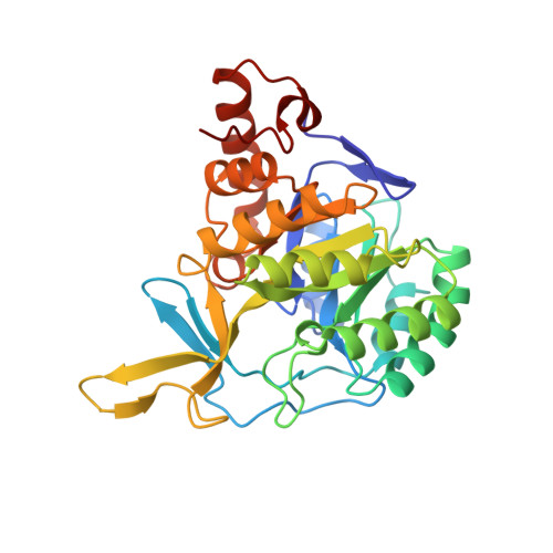
    Trypanosoma cruzi is the etiological agent of Chagas' disease, a pathogenesis that affects millions of people in Latin America. Here, we report the crystal structure of dihydroorotate dehydrogenase (DHODH) from T. cruzi strain Y solved at 2.2A resolution. DHODH is a flavin mononucleotide containing enzyme, which catalyses the oxidation of l-dihydroorotate to orotate, the fourth step and only redox reaction in the de novo biosynthesis of pyrimidine nucleotides. Genetic studies have shown that DHODH is essential for T. cruzi survival, validating the idea that this enzyme can be considered an attractive target for the development of antichagasic drugs. In our work, a detailed analysis of T. cruzi DHODH crystal structure has allowed us to suggest potential sites to be further exploited for the design of highly specific inhibitors through the technology of structure-based drug design.


  • Organizational Affiliation

    Laboratório de Cristalografia de Proteínas, Departamento de Física e Química, Faculdade de Ciências Farmacêuticas de Ribeirão Preto, Universidade de São Paulo, Ribeirão Preto S.P. 14040-903, Brazil.


Macromolecules
Find similar proteins by:  (by identity cutoff)  |  3D Structure
Entity ID: 1
MoleculeChains Sequence LengthOrganismDetailsImage
Dihydroorotate dehydrogenase
A, B, C, D
312Trypanosoma cruziMutation(s): 1 
Gene Names: tcdhod2
UniProt
Find proteins for Q4D3W2 (Trypanosoma cruzi (strain CL Brener))
Explore Q4D3W2 
Go to UniProtKB:  Q4D3W2
Entity Groups  
Sequence Clusters30% Identity50% Identity70% Identity90% Identity95% Identity100% Identity
UniProt GroupQ4D3W2
Sequence Annotations
Expand
  • Reference Sequence
Small Molecules
Ligands 3 Unique
IDChains Name / Formula / InChI Key2D Diagram3D Interactions
FMN
Query on FMN

Download Ideal Coordinates CCD File 
F [auth A],
J [auth B],
M [auth C],
P [auth D]
FLAVIN MONONUCLEOTIDE
C17 H21 N4 O9 P
FVTCRASFADXXNN-SCRDCRAPSA-N
SO4
Query on SO4

Download Ideal Coordinates CCD File 
E [auth A],
I [auth B],
L [auth C],
O [auth D]
SULFATE ION
O4 S
QAOWNCQODCNURD-UHFFFAOYSA-L
GOL
Query on GOL

Download Ideal Coordinates CCD File 
G [auth A],
H [auth A],
K [auth B],
N [auth C],
Q [auth D]
GLYCEROL
C3 H8 O3
PEDCQBHIVMGVHV-UHFFFAOYSA-N
Experimental Data & Validation

Experimental Data

  • Method: X-RAY DIFFRACTION
  • Resolution: 2.20 Å
  • R-Value Free: 0.251 
  • R-Value Work: 0.206 
  • R-Value Observed: 0.207 
  • Space Group: P 21 21 21
Unit Cell:
Length ( Å )Angle ( ˚ )
a = 82.39α = 90
b = 123.92β = 90
c = 128.89γ = 90
Software Package:
Software NamePurpose
XSCALEdata scaling
MOLREPphasing
CNSrefinement
PDB_EXTRACTdata extraction
MAR345dtbdata collection
XDSdata reduction

Structure Validation

View Full Validation Report


Ligand Structure Quality Assessment 


Entry History 

Deposition Data

Revision History  (Full details and data files)

  • Version 1.0: 2008-04-29
    Type: Initial release
  • Version 1.1: 2011-07-13
    Changes: Non-polymer description, Version format compliance
  • Version 1.2: 2024-02-21
    Changes: Data collection, Database references, Derived calculations