Structural studies of langerin and Birbeck granule: a macromolecular organization model
Thepaut, M., Valladeau, J., Nurisso, A., Kahn, R., Arnou, B., Vives, C., Saeland, S., Ebel, C., Monnier, C., Dezutter-Dambuyant, C., Imberty, A., Fieschi, F.(2009) Biochemistry 48: 2684-2698
- PubMed: 19175323
- DOI: https://doi.org/10.1021/bi802151w
- Primary Citation of Related Structures:
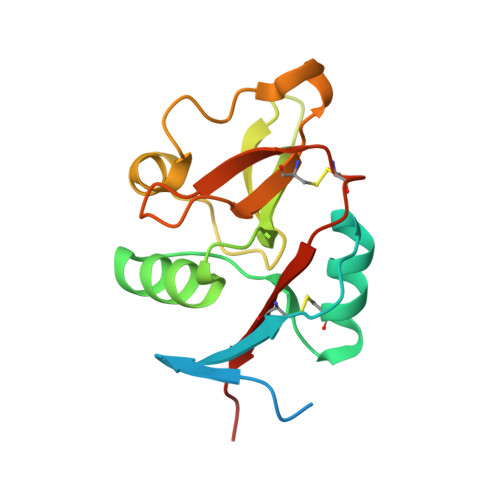
3C22 - PubMed Abstract:
Dendritic cells, a sentinel immunity cell lineage, include different cell subsets that express various C-type lectins. For example, epidermal Langerhans cells express langerin, and some dermal dendritic cells express DC-SIGN. Langerin is a crucial component of Birbeck granules, the Langerhans cell hallmark organelle, and may have a preventive role toward HIV, by its internalization into Birbeck granules. Since langerin carbohydrate recognition domain (CRD) is crucial for HIV interaction and Birbeck granule formation, we produced the CRD of human langerin and solved its structure at 1.5 A resolution. On this basis gp120 high-mannose oligosaccharide binding has been evaluated by molecular modeling. Hydrodynamic studies reveal a very elongated shape of recombinant langerin extracellular domain (ECD). A molecular model of the langerin ECD, integrating the CRD structure, has been generated and validated by comparison with hydrodynamic parameters. In parallel, Langerhans cells were isolated from human skin. From their analysis by electron microscopy and the langerin ECD model, an ultrastructural organization is proposed for Birbeck granules. To delineate the role of the different langerin domains in Birbeck granule formation, we generated truncated and mutated langerin constructs. After transfection into a fibroblastic cell line, we highlighted, in accordance with our model, the role of the CRD in the membrane zipping occurring in BG formation as well as some contribution of the cytoplasmic domain. Finally, we have shown that langerin ECD triggering with a specific mAb promotes global rearrangements of LC morphology. Our results open the way to the definition of a new membrane deformation mechanism.
Organizational Affiliation:
Laboratoire des Proteines Membranaires, CEA, DSV, Institut de Biologie Structurale (IBS), Grenoble, France.