


Crystal structure of spermidine synthase from Trypanosoma cruzi.
Bosch, J., Hol, W.G.J.To be published.
Experimental Data Snapshot
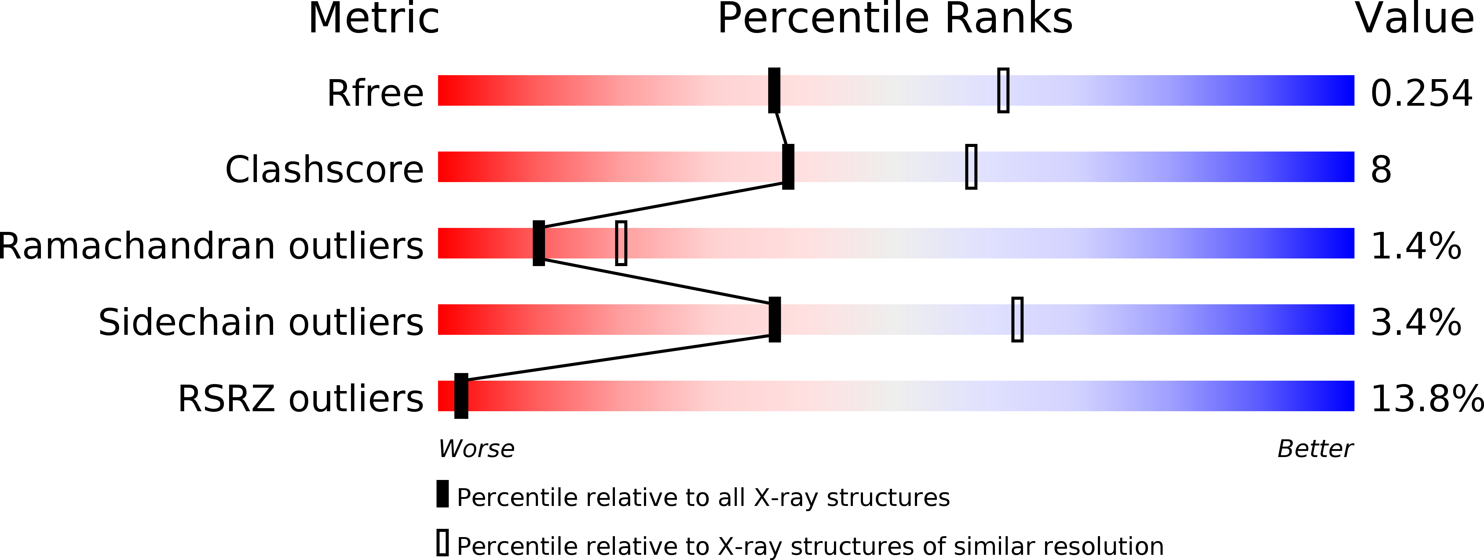
wwPDB Validation 3D Report Full Report
Entity ID: 1 | |||||
|---|---|---|---|---|---|
| Molecule | Chains | Sequence Length | Organism | Details | Image |
| Spermidine synthase | 304 | Trypanosoma cruzi strain CL Brener | Mutation(s): 0 Gene Names: Tc00.1047053510339.50 EC: 2.5.1.16 | 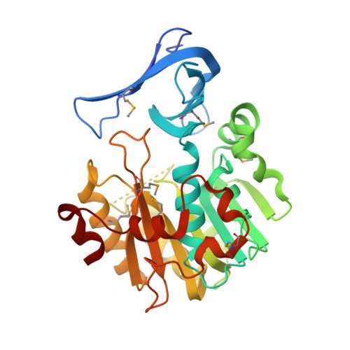 | |
UniProt | |||||
Find proteins for Q4DA73 (Trypanosoma cruzi (strain CL Brener)) Explore Q4DA73 Go to UniProtKB: Q4DA73 | |||||
Entity Groups | |||||
| Sequence Clusters | 30% Identity50% Identity70% Identity90% Identity95% Identity100% Identity | ||||
| UniProt Group | Q4DA73 | ||||
Sequence AnnotationsExpand | |||||
| |||||
| Modified Residues 1 Unique | |||||
|---|---|---|---|---|---|
| ID | Chains | Type | Formula | 2D Diagram | Parent |
| MSE Query on MSE | A, B | L-PEPTIDE LINKING | C5 H11 N O2 Se |  | MET |
| Length ( Å ) | Angle ( ˚ ) |
|---|---|
| a = 43.669 | α = 90 |
| b = 96.996 | β = 106.33 |
| c = 69.649 | γ = 90 |
| Software Name | Purpose |
|---|---|
| REFMAC | refinement |
| ADSC | data collection |
| XDS | data reduction |
| XSCALE | data scaling |
| SHELXD | phasing |
RCSB PDB (citation) is hosted by
RCSB PDB is a member of the

