3BVH

Crystal Structure of Recombinant gammaD364A Fibrinogen Fragment D with the Peptide Ligand Gly-Pro-Arg-Pro-Amide


Experimental Data Snapshot

  • Method: X-RAY DIFFRACTION
  • Resolution: 2.60 Å
  • R-Value Free: 0.260 
  • R-Value Work: 0.220 
  • R-Value Observed: 0.220 

wwPDB Validation   3D Report Full Report


This is version 2.2 of the entry. See complete history


Literature

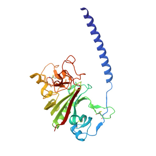
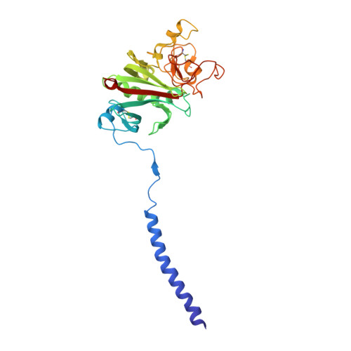
Polymerization-defective fibrinogen variant gammaD364A binds knob "A" peptide mimic.

Bowley, S.R.Merenbloom, B.K.Okumura, N.Betts, L.Heroux, A.Gorkun, O.V.Lord, S.T.

(2008) Biochemistry 47: 8607-8613

  • DOI: https://doi.org/10.1021/bi8000769
  • Primary Citation of Related Structures:  
    3BVH

  • PubMed Abstract: 

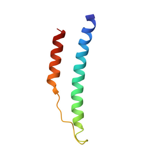
    Fibrin polymerization is supported in part by interactions called "A:a". Crystallographic studies revealed gamma364Asp is part of hole "a" that interacts with knob "A" peptide mimic, GPRP. Biochemical studies have shown gamma364Asp is critical to polymerization, as polymerization of variants gammaD364A, gammaD364H, and gammaD364V is exceptionally impaired. To understand the molecular basis for the aberrant function, we solved the crystal structure of fragment D from gammaD364A. Surprisingly, the structure (rfD-gammaD364A+GP) showed near normal "A:a" interactions with GPRP bound to hole "a" and no change in the overall structure of gammaD364A. Of note, inspection of the structure showed negative electrostatic potential inside hole "a" was diminished by this substitution. We examined GPRP binding to the gamma364Asp variants in solution by plasmin protection assay. We found no protection of either gammaD364H or gammaD364V but partial protection of gammaD364A, indicating the peptide does not bind to either gammaD364H or gammaD364V and binds more weakly than normal to gammaD364A. We also examined protection by calcium and found all variants were indistinguishable from normal, suggesting the global structures of the variants are not markedly different from normal. Our data imply that gamma364Asp per se is not required for knob "A" binding to hole "a"; rather, this residue's negative charge has a critical role in the electrostatic interactions that facilitate the important first step in fibrin polymerization.


  • Organizational Affiliation

    Department of Chemistry, University of North Carolina, Chapel Hill, North Carolina 27599, USA.


Macromolecules
Find similar proteins by:  (by identity cutoff)  |  3D Structure
Entity ID: 1
MoleculeChains Sequence LengthOrganismDetailsImage
Fibrinogen alpha chain
A, D
62Homo sapiensMutation(s): 0 
Gene Names: FGA
UniProt & NIH Common Fund Data Resources
Find proteins for P02671 (Homo sapiens)
Explore P02671 
Go to UniProtKB:  P02671
PHAROS:  P02671
GTEx:  ENSG00000171560 
Entity Groups  
Sequence Clusters30% Identity50% Identity70% Identity90% Identity95% Identity100% Identity
UniProt GroupP02671
Sequence Annotations
Expand
  • Reference Sequence
Find similar proteins by:  (by identity cutoff)  |  3D Structure
Entity ID: 2
MoleculeChains Sequence LengthOrganismDetailsImage
Fibrinogen beta chain
B, E
298Homo sapiensMutation(s): 0 
Gene Names: FGB
UniProt & NIH Common Fund Data Resources
Find proteins for P02675 (Homo sapiens)
Explore P02675 
Go to UniProtKB:  P02675
PHAROS:  P02675
GTEx:  ENSG00000171564 
Entity Groups  
Sequence Clusters30% Identity50% Identity70% Identity90% Identity95% Identity100% Identity
UniProt GroupP02675
Sequence Annotations
Expand
  • Reference Sequence
Find similar proteins by:  (by identity cutoff)  |  3D Structure
Entity ID: 3
MoleculeChains Sequence LengthOrganismDetailsImage
Fibrinogen gamma chain
C, F
293Homo sapiensMutation(s): 1 
Gene Names: FGG
UniProt & NIH Common Fund Data Resources
Find proteins for P02679 (Homo sapiens)
Explore P02679 
Go to UniProtKB:  P02679
PHAROS:  P02679
GTEx:  ENSG00000171557 
Entity Groups  
Sequence Clusters30% Identity50% Identity70% Identity90% Identity95% Identity100% Identity
UniProt GroupP02679
Sequence Annotations
Expand
  • Reference Sequence

Find similar proteins by:  Sequence   |   3D Structure  

Entity ID: 4
MoleculeChains Sequence LengthOrganismDetailsImage
4-mer peptide GPRPG,
H [auth I],
I [auth H],
J
4synthetic constructMutation(s): 0 
Sequence Annotations
Expand
  • Reference Sequence
Oligosaccharides

Help

Entity ID: 5
MoleculeChains Length2D Diagram Glycosylation3D Interactions
2-acetamido-2-deoxy-beta-D-glucopyranose-(1-4)-[alpha-L-fucopyranose-(1-6)]2-acetamido-2-deoxy-beta-D-glucopyranose
K, L
3N-Glycosylation
Glycosylation Resources
GlyTouCan:  G21290RB
GlyCosmos:  G21290RB
GlyGen:  G21290RB
Experimental Data & Validation

Experimental Data

  • Method: X-RAY DIFFRACTION
  • Resolution: 2.60 Å
  • R-Value Free: 0.260 
  • R-Value Work: 0.220 
  • R-Value Observed: 0.220 
  • Space Group: P 21 21 21
Unit Cell:
Length ( Å )Angle ( ˚ )
a = 89.844α = 90
b = 94.913β = 90
c = 225.754γ = 90
Software Package:
Software NamePurpose
HKL-2000data collection
CNSrefinement
DENZOdata reduction
SCALEPACKdata scaling
CNSphasing

Structure Validation

View Full Validation Report


Entry History 

Deposition Data

Revision History  (Full details and data files)

  • Version 1.0: 2008-09-02
    Type: Initial release
  • Version 1.1: 2011-07-13
    Changes: Non-polymer description, Version format compliance
  • Version 1.2: 2018-12-26
    Changes: Data collection, Derived calculations, Source and taxonomy
  • Version 2.0: 2020-07-29
    Type: Remediation
    Reason: Carbohydrate remediation
    Changes: Atomic model, Data collection, Derived calculations, Structure summary
  • Version 2.1: 2021-10-20
    Changes: Database references, Structure summary
  • Version 2.2: 2023-08-30
    Changes: Data collection, Refinement description