3BSE

Crystal structure analysis of a 16-base-pair B-DNA

  • Classification: DNA
  • Mutation(s): No 

  • Deposited: 2007-12-23 Released: 2008-12-23 
  • Deposition Author(s): Narayana, N.

Experimental Data Snapshot

  • Method: X-RAY DIFFRACTION
  • Resolution: 1.60 Å
  • R-Value Free: 0.274 
  • R-Value Observed: 0.221 

wwPDB Validation   3D Report Full Report


This is version 1.2 of the entry. See complete history


Literature

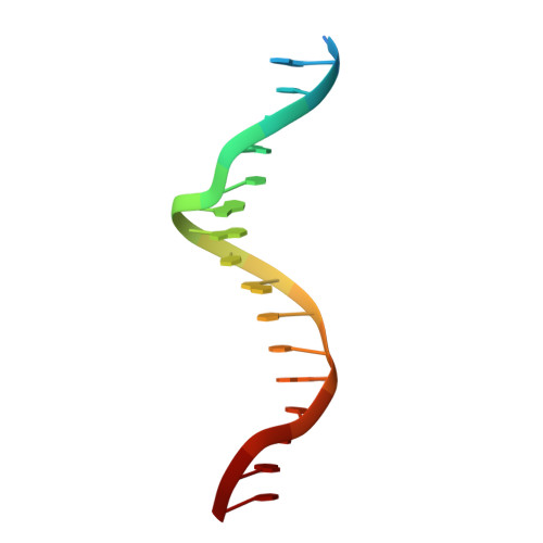
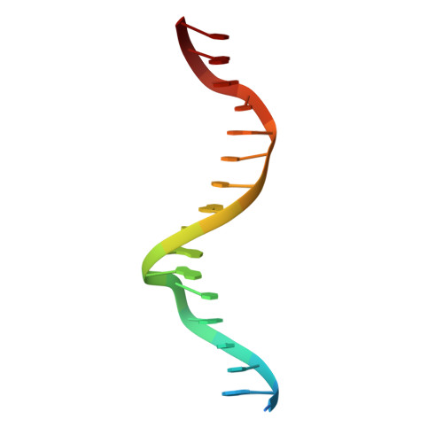
Crystallographic Analysis of a Sex-Specific Enhancer Element: Sequence-Dependent DNA Structure, Hydration, and Dynamics

Narayana, N.Weiss, M.A.

(2009) J Mol Biol 385: 469-490

  • DOI: https://doi.org/10.1016/j.jmb.2008.10.041
  • Primary Citation of Related Structures:  
    3BSE

  • PubMed Abstract: 

    The crystal structure of a sex-specific enhancer element is described at a resolution of 1.6 A. This 16-bp site, designated Dsx(A), functions in the regulation of a genetic switch between male and female patterns of gene expression in Drosophila melanogaster. Related sites are broadly conserved in metazoans, including in the human genome. This enhancer element is unusually rich in general regulatory sequences related to DNA recognition by multiple classes of eukaryotic transcription factors, including the DM motifs, homeodomain, and high mobility group box. Whereas free DNA is often crystallized as an A-form double helix, Dsx(A) was crystallized as B-DNA and thus provides a model for the prebound conformation of diverse regulatory DNA complexes. Sequence-dependent conformational properties that extend features of shorter B-DNA fragments with respect to double helical parameters, groove widths, hydration, and binding of divalent metal ions are observed. The structure also exhibits a sequence-dependent pattern of isotropic thermal B-factors, suggesting possible variation in the local flexibility of the DNA backbone. Such fluctuations are in accord with structural variability observed in prior B-DNA structures. We speculate that sites of intrinsic flexibility within a DNA control element provide hinges for its protein-directed reorganization in a transcriptional preinitiation complex.


  • Organizational Affiliation

    Department of Biochemistry, Case Western Reserve University, Cleveland, OH 44106, USA. narayanan3@uthscsa.edu


Macromolecules

Find similar nucleic acids by:  Sequence   |   3D Structure  

Entity ID: 1
MoleculeChains LengthOrganismImage
DNA (5'-D(*DA*DCP*DAP*DCP*DTP*DAP*DCP*DAP*DAP*DTP*DGP*DTP*DTP*DGP*DCP*DAP*DAP*DT)-3')18N/A
Sequence Annotations
Expand
  • Reference Sequence

Find similar nucleic acids by:  Sequence   |   3D Structure  

Entity ID: 2
MoleculeChains LengthOrganismImage
DNA (5'-D(*DG*DTP*DAP*DTP*DTP*DGP*DCP*DAP*DAP*DCP*DAP*DTP*DTP*DGP*DTP*DAP*DGP*DT)-3')18N/A
Sequence Annotations
Expand
  • Reference Sequence
Small Molecules
Ligands 1 Unique
IDChains Name / Formula / InChI Key2D Diagram3D Interactions
CA
Query on CA

Download Ideal Coordinates CCD File 
C [auth A]
D [auth A]
E [auth A]
F [auth A]
G [auth A]
C [auth A],
D [auth A],
E [auth A],
F [auth A],
G [auth A],
H [auth A],
I [auth A],
J [auth B],
K [auth B],
L [auth B],
M [auth B],
N [auth B]
CALCIUM ION
Ca
BHPQYMZQTOCNFJ-UHFFFAOYSA-N
Experimental Data & Validation

Experimental Data

  • Method: X-RAY DIFFRACTION
  • Resolution: 1.60 Å
  • R-Value Free: 0.274 
  • R-Value Observed: 0.221 
  • Space Group: H 3
Unit Cell:
Length ( Å )Angle ( ˚ )
a = 38.74α = 90
b = 38.74β = 90
c = 161.327γ = 120
Software Package:
Software NamePurpose
HKL-2000data collection
SOLVEphasing
CNSrefinement
HKL-2000data reduction
HKL-2000data scaling

Structure Validation

View Full Validation Report


Entry History 

Deposition Data

  • Released Date: 2008-12-23 
  • Deposition Author(s): Narayana, N.

Revision History  (Full details and data files)

  • Version 1.0: 2008-12-23
    Type: Initial release
  • Version 1.1: 2011-07-13
    Changes: Version format compliance
  • Version 1.2: 2024-02-21
    Changes: Data collection, Database references, Derived calculations