3BQU

Crystal Structure of the 2F5 Fab'-3H6 Fab Complex


Experimental Data Snapshot

  • Method: X-RAY DIFFRACTION
  • Resolution: 3.00 Å
  • R-Value Free: 0.295 
  • R-Value Work: 0.276 
  • R-Value Observed: 0.276 

wwPDB Validation   3D Report Full Report


This is version 1.2 of the entry. See complete history


Literature

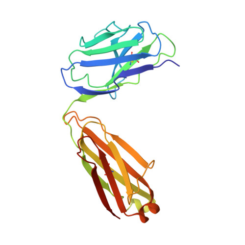
Crystal structure of the complex between the F(ab)' fragment of the cross-neutralizing anti-HIV-1 antibody 2F5 and the F(ab) fragment of its anti-idiotypic antibody 3H6.

Bryson, S.Julien, J.P.Isenman, D.E.Kunert, R.Katinger, H.Pai, E.F.

(2008) J Mol Biol 382: 910-919

  • DOI: https://doi.org/10.1016/j.jmb.2008.07.057
  • Primary Citation of Related Structures:  
    3BQU

  • PubMed Abstract: 

    The monoclonal antibody 2F5 neutralizes a broad range of human immunodeficiency virus-1 isolates via a conserved epitope on the viral glycoprotein gp41. The conformation of the principal epitope is a type I beta-turn centered on gp41 residues (664)DKW(666); in addition, binding studies indicate that residues N- and C-terminal to this core as well as structurally more distant parts of gp41 also contribute to the interaction. Ab2/3H6 is an anti-idiotypic antibody that inhibits the interaction between 2F5 and gp41 and as such, Ab2/3H6 may, in principle, possess a paratope that mimics the gp41 epitope. To establish the potential of Ab2/3H6 to serve as a guide for the design of vaccine components against human immunodeficiency virus, we investigated the crystal structure of the heterodimeric complex of Ab2/3H6 F(ab) and 2F5 F(ab)'. Ab2/3H6 F(ab) binds to 2F5 F(ab)' via a helix-like protrusion formed by residues (58(H))RYSPSLNTRL(67(H)) of the 2F5 F(ab)' variable domain and proximal to but not overlapping with the gp41 (664)DKW(666) epitope-binding pocket. This defines Ab2/3H6 as an anti-idiotypic antibody of the Ab2gamma class, i.e., an antigen-inhibitable idiotype that does not carry the internal image of the linear primary gp41 (662)ELDKWAS(668) epitope.


  • Organizational Affiliation

    Department of Biochemistry, University of Toronto, 1 King's College Circle, Toronto, Ontario, Canada M5S 1A8.


Macromolecules
Find similar proteins by:  (by identity cutoff)  |  3D Structure
Entity ID: 1
MoleculeChains Sequence LengthOrganismDetailsImage
2F5 Fab' light chain214Homo sapiensMutation(s): 0 
Entity Groups  
Sequence Clusters30% Identity50% Identity70% Identity90% Identity95% Identity100% Identity
Sequence Annotations
Expand
  • Reference Sequence
Find similar proteins by:  (by identity cutoff)  |  3D Structure
Entity ID: 2
MoleculeChains Sequence LengthOrganismDetailsImage
2F5 Fab' heavy chain235Homo sapiensMutation(s): 0 
UniProt
Find proteins for Q6PYX1 (Homo sapiens)
Explore Q6PYX1 
Go to UniProtKB:  Q6PYX1
Entity Groups  
Sequence Clusters30% Identity50% Identity70% Identity90% Identity95% Identity100% Identity
UniProt GroupQ6PYX1
Sequence Annotations
Expand
  • Reference Sequence
Find similar proteins by:  (by identity cutoff)  |  3D Structure
Entity ID: 3
MoleculeChains Sequence LengthOrganismDetailsImage
3H6 Fab light chain233Mus musculusMutation(s): 0 
UniProt
Find proteins for A2NHM3 (Mus musculus)
Explore A2NHM3 
Go to UniProtKB:  A2NHM3
Entity Groups  
Sequence Clusters30% Identity50% Identity70% Identity90% Identity95% Identity100% Identity
UniProt GroupA2NHM3
Sequence Annotations
Expand
  • Reference Sequence
Find similar proteins by:  (by identity cutoff)  |  3D Structure
Entity ID: 4
MoleculeChains Sequence LengthOrganismDetailsImage
3H6 Fab heavy chain241Mus musculusMutation(s): 0 
UniProt
Find proteins for P01756 (Mus musculus)
Explore P01756 
Go to UniProtKB:  P01756
Entity Groups  
Sequence Clusters30% Identity50% Identity70% Identity90% Identity95% Identity100% Identity
UniProt GroupP01756
Sequence Annotations
Expand
  • Reference Sequence
Small Molecules
Ligands 1 Unique
IDChains Name / Formula / InChI Key2D Diagram3D Interactions
SO4
Query on SO4

Download Ideal Coordinates CCD File 
E [auth D]SULFATE ION
O4 S
QAOWNCQODCNURD-UHFFFAOYSA-L
Experimental Data & Validation

Experimental Data

  • Method: X-RAY DIFFRACTION
  • Resolution: 3.00 Å
  • R-Value Free: 0.295 
  • R-Value Work: 0.276 
  • R-Value Observed: 0.276 
  • Space Group: P 21 21 21
Unit Cell:
Length ( Å )Angle ( ˚ )
a = 67.44α = 90
b = 98.33β = 90
c = 154.39γ = 90
Software Package:
Software NamePurpose
MAR345data collection
CNSrefinement
DENZOdata reduction
SCALEPACKdata scaling
CNSphasing

Structure Validation

View Full Validation Report


Entry History 

Deposition Data

Revision History  (Full details and data files)

  • Version 1.0: 2008-10-21
    Type: Initial release
  • Version 1.1: 2011-07-13
    Changes: Version format compliance
  • Version 1.2: 2023-08-30
    Changes: Data collection, Database references, Derived calculations, Refinement description