3BO8

The High Resolution Crystal Structure of HLA-A1 Complexed with the MAGE-A1 Peptide


Experimental Data Snapshot

  • Method: X-RAY DIFFRACTION
  • Resolution: 1.80 Å
  • R-Value Free: 0.225 
  • R-Value Work: 0.195 
  • R-Value Observed: 0.196 

wwPDB Validation   3D Report Full Report


This is version 1.2 of the entry. See complete history


Literature

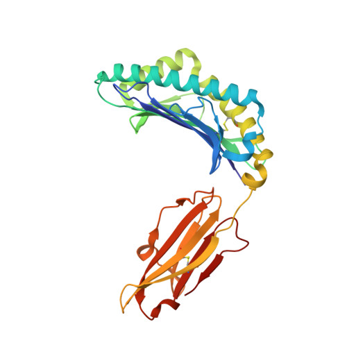
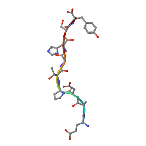
Conformational changes within the HLA-A1:MAGE-A1 complex induced by binding of a recombinant antibody fragment with TCR-like specificity

Kumar, P.Vahedi-Faridi, A.Saenger, W.Ziegler, A.Uchanska-Ziegler, B.

(2009) Protein Sci 18: 37-49

  • DOI: https://doi.org/10.1002/pro.4
  • Primary Citation of Related Structures:  
    3BO8

  • PubMed Abstract: 

    Although there is X-ray crystallographic evidence that the interaction between major histocompatibility complex (MHC, in humans HLA) class I molecules and T cell receptors (TCR) or killer cell Ig-like receptors (KIR) may be accompanied by considerable changes in the conformation of selected residues or even entire loops within TCR or KIR, conformational changes between receptor-bound and -unbound MHC class I molecules of comparable magnitude have not been observed so far. We have previously determined the structure of the MHC class I molecule HLA-A1 bound to a melanoma antigen-encoding gene (MAGE)-A1-derived peptide in complex with a recombinant antibody fragment with TCR-like specificity, Fab-Hyb3. Here, we compare the X-ray structure of HLA-A1:MAGE-A1 with that complexed with Fab-Hyb3 to gain insight into structural changes of the MHC molecule that might be induced by the interaction with the antibody fragment. Apart from the expulsion of several water molecules from the interface, Fab-Hyb3 binding results in major rearrangements (up to 5.5 A) of heavy chain residues Arg65, Gln72, Arg145, and Lys146. Residue 65 is frequently and residues 72 and 146 are occasionally involved in TCR binding-induced conformational changes, as revealed by a comparison with MHC class I structures in TCR-liganded and -unliganded forms. On the other hand, residue 145 is subject to a reorientation following engagement of HLA-Cw4 and KIR2DL1. Therefore, conformational changes within the HLA-A1:MAGE-A1:Fab-Hyb3 complex include MHC residues that are also involved in reorientations in complexes with natural ligands, pointing to their central importance for the peptide-dependent recognition of MHC molecules.


  • Organizational Affiliation

    Institut für Immungenetik, Charité-Universitätsmedizin Berlin, Campus Benjamin Franklin, Freie Universität Berlin, Thielallee 73, Berlin 14195, Germany.


Macromolecules
Find similar proteins by:  (by identity cutoff)  |  3D Structure
Entity ID: 1
MoleculeChains Sequence LengthOrganismDetailsImage
HLA class I histocompatibility antigen, A-1 alpha chain274Homo sapiensMutation(s): 0 
UniProt & NIH Common Fund Data Resources
Find proteins for P04439 (Homo sapiens)
Explore P04439 
Go to UniProtKB:  P04439
PHAROS:  P04439
GTEx:  ENSG00000206503 
Entity Groups  
Sequence Clusters30% Identity50% Identity70% Identity90% Identity95% Identity100% Identity
UniProt GroupP04439
Sequence Annotations
Expand
  • Reference Sequence
Find similar proteins by:  (by identity cutoff)  |  3D Structure
Entity ID: 2
MoleculeChains Sequence LengthOrganismDetailsImage
Beta-2-microglobulin100Homo sapiensMutation(s): 0 
UniProt & NIH Common Fund Data Resources
Find proteins for P61769 (Homo sapiens)
Explore P61769 
Go to UniProtKB:  P61769
PHAROS:  P61769
GTEx:  ENSG00000166710 
Entity Groups  
Sequence Clusters30% Identity50% Identity70% Identity90% Identity95% Identity100% Identity
UniProt GroupP61769
Sequence Annotations
Expand
  • Reference Sequence

Find similar proteins by:  Sequence   |   3D Structure  

Entity ID: 3
MoleculeChains Sequence LengthOrganismDetailsImage
nonameric peptide from Melanoma-associated antigen 19N/AMutation(s): 0 
UniProt & NIH Common Fund Data Resources
Find proteins for P43355 (Homo sapiens)
Explore P43355 
Go to UniProtKB:  P43355
PHAROS:  P43355
GTEx:  ENSG00000198681 
Entity Groups  
UniProt GroupP43355
Sequence Annotations
Expand
  • Reference Sequence
Experimental Data & Validation

Experimental Data

  • Method: X-RAY DIFFRACTION
  • Resolution: 1.80 Å
  • R-Value Free: 0.225 
  • R-Value Work: 0.195 
  • R-Value Observed: 0.196 
  • Space Group: P 21 21 21
Unit Cell:
Length ( Å )Angle ( ˚ )
a = 51.176α = 90
b = 74.06β = 90
c = 125.94γ = 90
Software Package:
Software NamePurpose
MOSFLMdata reduction
SCALAdata scaling
MOLREPphasing
REFMACrefinement
PDB_EXTRACTdata extraction
ADSCdata collection

Structure Validation

View Full Validation Report


Entry History 

Deposition Data

Revision History  (Full details and data files)

  • Version 1.0: 2008-12-23
    Type: Initial release
  • Version 1.1: 2011-07-13
    Changes: Non-polymer description, Version format compliance
  • Version 1.2: 2023-11-01
    Changes: Data collection, Database references, Derived calculations, Refinement description