3BDJ

Crystal Structure of Bovine Milk Xanthine Dehydrogenase with a Covalently Bound Oxipurinol Inhibitor


Experimental Data Snapshot

  • Method: X-RAY DIFFRACTION
  • Resolution: 2.00 Å
  • R-Value Free: 0.224 
  • R-Value Work: 0.191 
  • R-Value Observed: 0.191 

wwPDB Validation   3D Report Full Report


Ligand Structure Quality Assessment 


This is version 1.3 of the entry. See complete history


Literature

Mechanism of inhibition of xanthine oxidoreductase by allopurinol: crystal structure of reduced bovine milk xanthine oxidoreductase bound with oxipurinol.

Okamoto, K.Eger, B.T.Nishino, T.Pai, E.F.Nishino, T.

(2008) Nucleosides Nucleotides Nucleic Acids 27: 888-893

  • DOI: https://doi.org/10.1080/15257770802146577
  • Primary Citation of Related Structures:  
    3BDJ

  • PubMed Abstract: 

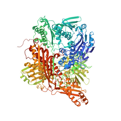
    Inhibitors of xanthine oxidoreductase block conversion of xanthine to uric acid and are therefore potentially useful for treatment of hyperuricemia or gout. We determined the crystal structure of reduced bovine milk xanthine oxidoreductase complexed with oxipurinol at 2.0 A resolution. Clear electron density was observed between the N2 nitrogen of oxipurinol and the molybdenum atom of the molybdopterin cofactor, indicating that oxipurinol coordinated directly to molybdenum. Oxipurinol forms hydrogen bonds with glutamate 802, arginine 880, and glutamate 1261, which have previously been shown to be essential for the enzyme reaction. We discuss possible differences in the hypouricemic effect of inhibitors, including allopurinol and newly developed inhibitors, based on their mode of binding in the crystal structures.


  • Organizational Affiliation

    Department of Biochemistry and Molecular Biology, Nippon Medical School, Tokyo, Japan.


Macromolecules
Find similar proteins by:  (by identity cutoff)  |  3D Structure
Entity ID: 1
MoleculeChains Sequence LengthOrganismDetailsImage
Xanthine dehydrogenase/oxidase
A, B
1,332Bos taurusMutation(s): 0 
EC: 1.17.3.2 (PDB Primary Data), 1.17.1.4 (PDB Primary Data)
UniProt
Find proteins for P80457 (Bos taurus)
Explore P80457 
Go to UniProtKB:  P80457
Entity Groups  
Sequence Clusters30% Identity50% Identity70% Identity90% Identity95% Identity100% Identity
UniProt GroupP80457
Sequence Annotations
Expand
  • Reference Sequence
Small Molecules
Ligands 8 Unique
IDChains Name / Formula / InChI Key2D Diagram3D Interactions
FAD
Query on FAD

Download Ideal Coordinates CCD File 
H [auth A],
S [auth B]
FLAVIN-ADENINE DINUCLEOTIDE
C27 H33 N9 O15 P2
VWWQXMAJTJZDQX-UYBVJOGSSA-N
MTE
Query on MTE

Download Ideal Coordinates CCD File 
F [auth A],
Q [auth B]
PHOSPHONIC ACIDMONO-(2-AMINO-5,6-DIMERCAPTO-4-OXO-3,7,8A,9,10,10A-HEXAHYDRO-4H-8-OXA-1,3,9,10-TETRAAZA-ANTHRACEN-7-YLMETHYL)ESTER
C10 H14 N5 O6 P S2
HPEUEJRPDGMIMY-IFQPEPLCSA-N
FES
Query on FES

Download Ideal Coordinates CCD File 
D [auth A],
E [auth A],
O [auth B],
P [auth B]
FE2/S2 (INORGANIC) CLUSTER
Fe2 S2
NIXDOXVAJZFRNF-UHFFFAOYSA-N
141
Query on 141

Download Ideal Coordinates CCD File 
I [auth A],
T [auth B]
Oxypurinol
C5 H4 N4 O2
HXNFUBHNUDHIGC-UHFFFAOYSA-N
MOW
Query on MOW

Download Ideal Coordinates CCD File 
G [auth A],
R [auth B]
Oxo(sulfanyl)molybdenum(IV) ION
H Mo O S
LCFBNOLMHXMMKH-UHFFFAOYSA-M
GOL
Query on GOL

Download Ideal Coordinates CCD File 
J [auth A]
K [auth A]
L [auth A]
U [auth B]
V [auth B]
J [auth A],
K [auth A],
L [auth A],
U [auth B],
V [auth B],
W [auth B],
X [auth B]
GLYCEROL
C3 H8 O3
PEDCQBHIVMGVHV-UHFFFAOYSA-N
CO3
Query on CO3

Download Ideal Coordinates CCD File 
M [auth A],
Y [auth B]
CARBONATE ION
C O3
BVKZGUZCCUSVTD-UHFFFAOYSA-L
CA
Query on CA

Download Ideal Coordinates CCD File 
C [auth A],
N [auth B]
CALCIUM ION
Ca
BHPQYMZQTOCNFJ-UHFFFAOYSA-N
Experimental Data & Validation

Experimental Data

  • Method: X-RAY DIFFRACTION
  • Resolution: 2.00 Å
  • R-Value Free: 0.224 
  • R-Value Work: 0.191 
  • R-Value Observed: 0.191 
  • Space Group: C 1 2 1
Unit Cell:
Length ( Å )Angle ( ˚ )
a = 166.517α = 90
b = 124.074β = 91.01
c = 148.797γ = 90
Software Package:
Software NamePurpose
ADSCdata collection
EPMRphasing
CNSrefinement
DENZOdata reduction
SCALEPACKdata scaling

Structure Validation

View Full Validation Report


Ligand Structure Quality Assessment 


Entry History 

Deposition Data

Revision History  (Full details and data files)

  • Version 1.0: 2008-11-25
    Type: Initial release
  • Version 1.1: 2011-07-13
    Changes: Non-polymer description, Version format compliance
  • Version 1.2: 2014-09-10
    Changes: Non-polymer description
  • Version 1.3: 2023-08-30
    Changes: Data collection, Database references, Derived calculations, Refinement description