3B4M

Crystal Structure of Human PABPN1 RRM


Experimental Data Snapshot

  • Method: X-RAY DIFFRACTION
  • Resolution: 2.82 Å
  • R-Value Free: 0.290 
  • R-Value Work: 0.226 
  • R-Value Observed: 0.229 

wwPDB Validation   3D Report Full Report


This is version 1.2 of the entry. See complete history


Literature

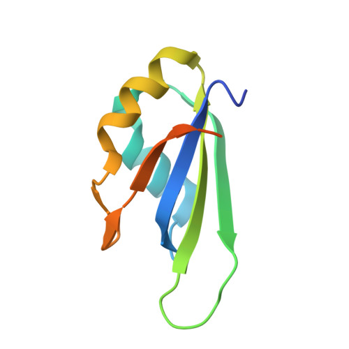
Crystal structure and possible dimerization of the single RRM of human PABPN1

Ge, H.Zhou, D.Tong, S.Gao, Y.Teng, M.Niu, L.

(2008) Proteins 71: 1539-1545


Macromolecules
Find similar proteins by:  (by identity cutoff)  |  3D Structure
Entity ID: 1
MoleculeChains Sequence LengthOrganismDetailsImage
Polyadenylate-binding protein 2
A, B, C, D
96Homo sapiensMutation(s): 0 
Gene Names: PABPN1PAB2PABP2
UniProt & NIH Common Fund Data Resources
Find proteins for Q86U42 (Homo sapiens)
Explore Q86U42 
Go to UniProtKB:  Q86U42
PHAROS:  Q86U42
GTEx:  ENSG00000100836 
Entity Groups  
Sequence Clusters30% Identity50% Identity70% Identity90% Identity95% Identity100% Identity
UniProt GroupQ86U42
Sequence Annotations
Expand
  • Reference Sequence
Experimental Data & Validation

Experimental Data

  • Method: X-RAY DIFFRACTION
  • Resolution: 2.82 Å
  • R-Value Free: 0.290 
  • R-Value Work: 0.226 
  • R-Value Observed: 0.229 
  • Space Group: P 31
Unit Cell:
Length ( Å )Angle ( ˚ )
a = 59.338α = 90
b = 59.338β = 90
c = 80.599γ = 120
Software Package:
Software NamePurpose
REFMACrefinement
AUTOMARdata reduction
MOLREPphasing

Structure Validation

View Full Validation Report


Entry History 

Deposition Data

Revision History  (Full details and data files)

  • Version 1.0: 2008-01-15
    Type: Initial release
  • Version 1.1: 2011-07-13
    Changes: Version format compliance
  • Version 1.2: 2023-11-01
    Changes: Data collection, Database references, Refinement description