Substrate tRNA recognition mechanism of a multisite-specific tRNA methyltransferase, Aquifex aeolicus Trm1, based on the X-ray crystal structure
Awai, T., Ochi, A., Ihsanawati, Sengoku, T., Hirata, A., Bessho, Y., Yokoyama, S., Hori, H.(2011) J Biol Chem 286: 35236-35246
- PubMed: 21844194
- DOI: https://doi.org/10.1074/jbc.M111.253641
- Primary Citation of Related Structures:
3AXS, 3AXT - PubMed Abstract:
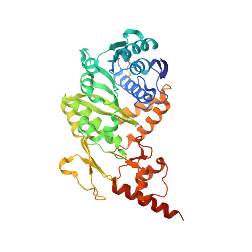
Archaeal and eukaryotic tRNA (N(2),N(2)-guanine)-dimethyltransferase (Trm1) produces N(2),N(2)-dimethylguanine at position 26 in tRNA. In contrast, Trm1 from Aquifex aeolicus, a hyper-thermophilic eubacterium, modifies G27 as well as G26. Here, a gel mobility shift assay revealed that the T-arm in tRNA is the binding site of A. aeolicus Trm1. To address the multisite specificity, we performed an x-ray crystal structure study. The overall structure of A. aeolicus Trm1 is similar to that of archaeal Trm1, although there is a zinc-cysteine cluster in the C-terminal domain of A. aeolicus Trm1. The N-terminal domain is a typical catalytic domain of S-adenosyl-l-methionine-dependent methyltransferases. On the basis of the crystal structure and amino acid sequence alignment, we prepared 30 mutant Trm1 proteins. These mutant proteins clarified residues important for S-adenosyl-l-methionine binding and enabled us to propose a hypothetical reaction mechanism. Furthermore, the tRNA-binding site was also elucidated by methyl transfer assay and gel mobility shift assay. The electrostatic potential surface models of A. aeolicus and archaeal Trm1 proteins demonstrated that the distribution of positive charges differs between the two proteins. We constructed a tRNA-docking model, in which the T-arm structure was placed onto the large area of positive charge, which is the expected tRNA-binding site, of A. aeolicus Trm1. In this model, the target G26 base can be placed near the catalytic pocket; however, the nucleotide at position 27 gains closer access to the pocket. Thus, this docking model introduces a rational explanation of the multisite specificity of A. aeolicus Trm1.
Organizational Affiliation:
Department of Materials Science and Biotechnology, Graduate School of Science and Engineering, Ehime University, Bunkyo 3, Matsuyama, Ehime 790-8577, Japan.