3ALQ

Crystal structure of TNF-TNFR2 complex


Experimental Data Snapshot

  • Method: X-RAY DIFFRACTION
  • Resolution: 3.00 Å
  • R-Value Free: 0.281 
  • R-Value Work: 0.213 
  • R-Value Observed: 0.220 

wwPDB Validation   3D Report Full Report


This is version 1.2 of the entry. See complete history


Literature

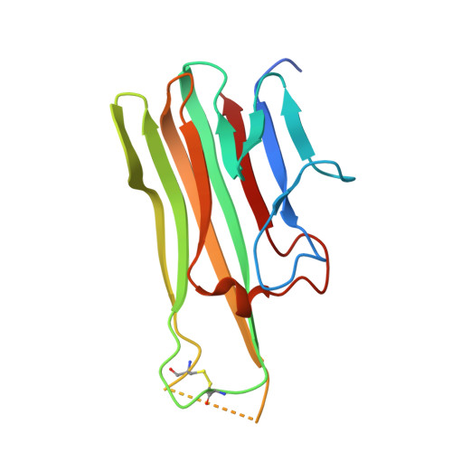
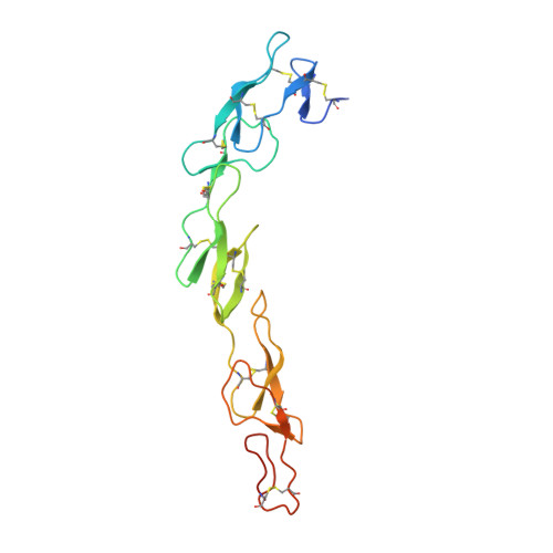
Solution of the Structure of the TNF-TNFR2 Complex

Mukai, Y.Nakamura, T.Yoshikawa, M.Yoshioka, Y.Tsunoda, S.I.Nakagawa, S.Yamagata, Y.Tsutsumi, Y.

(2010) Sci Signal 3: ra83-ra83

  • DOI: https://doi.org/10.1126/scisignal.2000954
  • Primary Citation of Related Structures:  
    3ALQ

  • PubMed Abstract: 

    Tumor necrosis factor (TNF) is an inflammatory cytokine that has important roles in various immune responses, which are mediated through its two receptors, TNF receptor 1 (TNFR1) and TNFR2. Antibody-based therapy against TNF is used clinically to treat several chronic autoimmune diseases; however, such treatment sometimes results in serious side effects, which are thought to be caused by the blocking of signals from both TNFRs. Therefore, knowledge of the structural basis for the recognition of TNF by each receptor would be invaluable in designing TNFR-selective drugs. Here, we solved the 3.0 angstrom resolution structure of the TNF-TNFR2 complex, which provided insight into the molecular recognition of TNF by TNFR2. Comparison to the known TNFR1 structure highlighted several differences between the ligand-binding interfaces of the two receptors. Additionally, we also demonstrated that TNF-TNFR2 formed aggregates on the surface of cells, which may be required for signal initiation. These results may contribute to the design of therapeutics for autoimmune diseases.


  • Organizational Affiliation

    Laboratory of Biopharmaceutical Research, National Institute of Biomedical Innovation, Osaka, Japan. yohe@phs.osaka-u.ac.jp


Macromolecules
Find similar proteins by:  (by identity cutoff)  |  3D Structure
Entity ID: 1
MoleculeChains Sequence LengthOrganismDetailsImage
Tumor necrosis factor
A, B, C, D, E
A, B, C, D, E, F
157Homo sapiensMutation(s): 6 
Gene Names: TNFTNFATNFSF2
UniProt & NIH Common Fund Data Resources
Find proteins for P01375 (Homo sapiens)
Explore P01375 
Go to UniProtKB:  P01375
PHAROS:  P01375
GTEx:  ENSG00000232810 
Entity Groups  
Sequence Clusters30% Identity50% Identity70% Identity90% Identity95% Identity100% Identity
UniProt GroupP01375
Sequence Annotations
Expand
  • Reference Sequence
Find similar proteins by:  (by identity cutoff)  |  3D Structure
Entity ID: 2
MoleculeChains Sequence LengthOrganismDetailsImage
Tumor necrosis factor receptor superfamily member 1B173Homo sapiensMutation(s): 0 
Gene Names: TNFBRTNFR2TNFRSF1B
UniProt & NIH Common Fund Data Resources
Find proteins for P20333 (Homo sapiens)
Explore P20333 
Go to UniProtKB:  P20333
PHAROS:  P20333
GTEx:  ENSG00000028137 
Entity Groups  
Sequence Clusters30% Identity50% Identity70% Identity90% Identity95% Identity100% Identity
UniProt GroupP20333
Sequence Annotations
Expand
  • Reference Sequence
Experimental Data & Validation

Experimental Data

  • Method: X-RAY DIFFRACTION
  • Resolution: 3.00 Å
  • R-Value Free: 0.281 
  • R-Value Work: 0.213 
  • R-Value Observed: 0.220 
  • Space Group: P 21 21 21
Unit Cell:
Length ( Å )Angle ( ˚ )
a = 74.47α = 90
b = 117.356β = 90
c = 246.835γ = 90
Software Package:
Software NamePurpose
HKL-2000data collection
AMoREphasing
PHENIXrefinement
HKL-2000data reduction
HKL-2000data scaling

Structure Validation

View Full Validation Report


Entry History 

Deposition Data

Revision History  (Full details and data files)

  • Version 1.0: 2010-11-17
    Type: Initial release
  • Version 1.1: 2011-07-13
    Changes: Version format compliance
  • Version 1.2: 2023-11-01
    Changes: Data collection, Database references, Derived calculations, Refinement description