3A9C

Crystal structure of ribose-1,5-bisphosphate isomerase from Thermococcus kodakaraensis KOD1 in complex with ribulose-1,5-bisphosphate


Experimental Data Snapshot

  • Method: X-RAY DIFFRACTION
  • Resolution: 2.60 Å
  • R-Value Free: 0.255 
  • R-Value Work: 0.201 
  • R-Value Observed: 0.201 

wwPDB Validation   3D Report Full Report


Ligand Structure Quality Assessment 


This is version 1.4 of the entry. See complete history


Literature

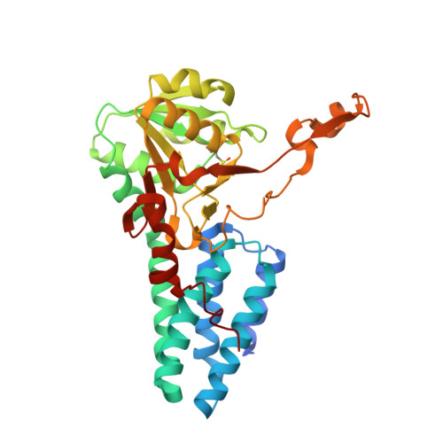
Dynamic, ligand-dependent conformational change triggers reaction of ribose-1,5-bisphosphate isomerase from Thermococcus kodakarensis KOD1

Nakamura, A.Fujihashi, M.Aono, R.Sato, T.Nishiba, Y.Yoshida, S.Yano, A.Atomi, H.Imanaka, T.Miki, K.

(2012) J Biol Chem 287: 20784-20796

  • DOI: https://doi.org/10.1074/jbc.M112.349423
  • Primary Citation of Related Structures:  
    3A11, 3A9C

  • PubMed Abstract: 

    Ribose-1,5-bisphosphate isomerase (R15Pi) is a novel enzyme recently identified as a member of an AMP metabolic pathway in archaea. The enzyme converts d-ribose 1,5-bisphosphate into ribulose 1,5-bisphosphate, providing the substrate for archaeal ribulose-1,5-bisphosphate carboxylase/oxygenases. We here report the crystal structures of R15Pi from Thermococcus kodakarensis KOD1 (Tk-R15Pi) with and without its substrate or product. Tk-R15Pi is a hexameric enzyme formed by the trimerization of dimer units. Biochemical analyses show that Tk-R15Pi only accepts the α-anomer of d-ribose 1,5-bisphosphate and that Cys(133) and Asp(202) residues are essential for ribulose 1,5-bisphosphate production. Comparison of the determined structures reveals that the unliganded and product-binding structures are in an open form, whereas the substrate-binding structure adopts a closed form, indicating domain movement upon substrate binding. The conformational change to the closed form optimizes active site configuration and also isolates the active site from the solvent, which may allow deprotonation of Cys(133) and protonation of Asp(202) to occur. The structural features of the substrate-binding form and biochemical evidence lead us to propose that the isomerase reaction proceeds via a cis-phosphoenolate intermediate.


  • Organizational Affiliation

    Department of Chemistry, Graduate School of Science, Kyoto University, Sakyo-ku, Kyoto 606-8502, Japan.


Macromolecules
Find similar proteins by:  (by identity cutoff)  |  3D Structure
Entity ID: 1
MoleculeChains Sequence LengthOrganismDetailsImage
Translation initiation factor eIF-2B, delta subunit
A, B, C, D, E
A, B, C, D, E, F
338Thermococcus kodakarensisMutation(s): 1 
Gene Names: E2b2
EC: 5.3.1
UniProt
Find proteins for Q5JFM9 (Thermococcus kodakarensis (strain ATCC BAA-918 / JCM 12380 / KOD1))
Explore Q5JFM9 
Go to UniProtKB:  Q5JFM9
Entity Groups  
Sequence Clusters30% Identity50% Identity70% Identity90% Identity95% Identity100% Identity
UniProt GroupQ5JFM9
Sequence Annotations
Expand
  • Reference Sequence
Small Molecules
Ligands 3 Unique
IDChains Name / Formula / InChI Key2D Diagram3D Interactions
RUB
Query on RUB

Download Ideal Coordinates CCD File 
G [auth A]
J [auth B]
M [auth C]
O [auth D]
Q [auth E]
G [auth A],
J [auth B],
M [auth C],
O [auth D],
Q [auth E],
S [auth F]
RIBULOSE-1,5-DIPHOSPHATE
C5 H12 O11 P2
YAHZABJORDUQGO-NQXXGFSBSA-N
PEG
Query on PEG

Download Ideal Coordinates CCD File 
H [auth A]
K [auth B]
N [auth C]
P [auth D]
R [auth E]
H [auth A],
K [auth B],
N [auth C],
P [auth D],
R [auth E],
T [auth F]
DI(HYDROXYETHYL)ETHER
C4 H10 O3
MTHSVFCYNBDYFN-UHFFFAOYSA-N
MG
Query on MG

Download Ideal Coordinates CCD File 
I [auth A],
L [auth B]
MAGNESIUM ION
Mg
JLVVSXFLKOJNIY-UHFFFAOYSA-N
Modified Residues  1 Unique
IDChains TypeFormula2D DiagramParent
CSD
Query on CSD
A, B, C, D, E
A, B, C, D, E, F
L-PEPTIDE LINKINGC3 H7 N O4 SCYS
Experimental Data & Validation

Experimental Data

  • Method: X-RAY DIFFRACTION
  • Resolution: 2.60 Å
  • R-Value Free: 0.255 
  • R-Value Work: 0.201 
  • R-Value Observed: 0.201 
  • Space Group: P 21 21 21
Unit Cell:
Length ( Å )Angle ( ˚ )
a = 117.313α = 90
b = 130.615β = 90
c = 132.672γ = 90
Software Package:
Software NamePurpose
CNSrefinement
HKL-2000data reduction
SCALEPACKdata scaling
CNSphasing

Structure Validation

View Full Validation Report


Ligand Structure Quality Assessment 


Entry History 

Deposition Data

Revision History  (Full details and data files)

  • Version 1.0: 2010-11-03
    Type: Initial release
  • Version 1.1: 2011-07-13
    Changes: Version format compliance
  • Version 1.2: 2012-05-02
    Changes: Database references
  • Version 1.3: 2013-06-12
    Changes: Database references
  • Version 1.4: 2023-11-01
    Changes: Data collection, Database references, Derived calculations, Refinement description