3A21

Crystal Structure of Streptomyces avermitilis beta-L-Arabinopyranosidase


Experimental Data Snapshot

  • Method: X-RAY DIFFRACTION
  • Resolution: 1.51 Å
  • R-Value Free: 0.179 
  • R-Value Work: 0.157 
  • R-Value Observed: 0.158 

wwPDB Validation   3D Report Full Report


This is version 1.2 of the entry. See complete history


Literature

A beta-l-Arabinopyranosidase from Streptomyces avermitilis is a novel member of glycoside hydrolase family 27.

Ichinose, H.Fujimoto, Z.Honda, M.Harazono, K.Nishimoto, Y.Uzura, A.Kaneko, S.

(2009) J Biol Chem 284: 25097-25106

  • DOI: https://doi.org/10.1074/jbc.M109.022723
  • Primary Citation of Related Structures:  
    3A21

  • PubMed Abstract: 

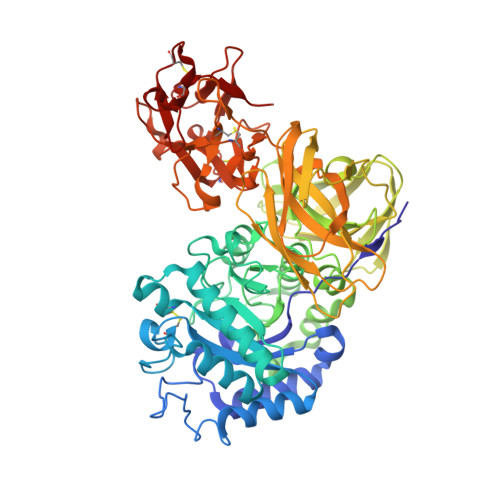
    Arabinogalactan proteins (AGPs) are a family of plant cell surface proteoglycans and are considered to be involved in plant growth and development. Because AGPs are very complex molecules, glycoside hydrolases capable of degrading AGPs are powerful tools for analyses of the AGPs. We previously reported such enzymes from Streptomyces avermitilis. Recently, a beta-l-arabinopyranosidase was purified from the culture supernatant of the bacterium, and its corresponding gene was identified. The primary structure of the protein revealed that the catalytic module was highly similar to that of glycoside hydrolase family 27 (GH27) alpha-d-galactosidases. The recombinant protein was successfully expressed as a secreted 64-kDa protein using a Streptomyces expression system. The specific activity toward p-nitrophenyl-beta-l-arabinopyranoside was 18 micromol of arabinose/min/mg, which was 67 times higher than that toward p- nitrophenyl-alpha-d-galactopyranoside. The enzyme could remove 0.1 and 45% l-arabinose from gum arabic or larch arabinogalactan, respectively. X-ray crystallographic analysis reveals that the protein had a GH27 catalytic domain, an antiparallel beta-domain containing Greek key motifs, another antiparallel beta-domain forming a jellyroll structure, and a carbohydrate-binding module family 13 domain. Comparison of the structure of this protein with that of alpha-d-galactosidase showed a single amino acid substitution (aspartic acid to glutamic acid) in the catalytic pocket of beta-l-arabinopyranosidase, and a space for the hydroxymethyl group on the C-5 carbon of d-galactose bound to alpha-galactosidase was changed in beta-l-arabinopyranosidase. Mutagenesis study revealed that the residue is critical for modulating the enzyme activity. This is the first report in which beta-l-arabinopyranosidase is classified as a new member of the GH27 family.


  • Organizational Affiliation

    Food Biotechnology Division, National Food Research Institute, 2-1-12 Kannondai, Tsukuba, Ibaraki 305-8642, Japan.


Macromolecules
Find similar proteins by:  (by identity cutoff)  |  3D Structure
Entity ID: 1
MoleculeChains Sequence LengthOrganismDetailsImage
Putative secreted alpha-galactosidase
A, B
614Streptomyces avermitilisMutation(s): 0 
Gene Names: agaA2SAV2186SAV_2186
UniProt
Find proteins for Q82L26 (Streptomyces avermitilis (strain ATCC 31267 / DSM 46492 / JCM 5070 / NBRC 14893 / NCIMB 12804 / NRRL 8165 / MA-4680))
Explore Q82L26 
Go to UniProtKB:  Q82L26
Entity Groups  
Sequence Clusters30% Identity50% Identity70% Identity90% Identity95% Identity100% Identity
UniProt GroupQ82L26
Sequence Annotations
Expand
  • Reference Sequence
Small Molecules
Ligands 4 Unique
IDChains Name / Formula / InChI Key2D Diagram3D Interactions
1PG
Query on 1PG

Download Ideal Coordinates CCD File 
JA [auth B],
KA [auth B],
O [auth A],
P [auth A]
2-(2-{2-[2-(2-METHOXY-ETHOXY)-ETHOXY]-ETHOXY}-ETHOXY)-ETHANOL
C11 H24 O6
SLNYBUIEAMRFSZ-UHFFFAOYSA-N
EPE
Query on EPE

Download Ideal Coordinates CCD File 
IA [auth B]4-(2-HYDROXYETHYL)-1-PIPERAZINE ETHANESULFONIC ACID
C8 H18 N2 O4 S
JKMHFZQWWAIEOD-UHFFFAOYSA-N
SO4
Query on SO4

Download Ideal Coordinates CCD File 
LA [auth B]
MA [auth B]
NA [auth B]
Q [auth A]
R [auth A]
LA [auth B],
MA [auth B],
NA [auth B],
Q [auth A],
R [auth A],
S [auth A],
T [auth A],
U [auth A]
SULFATE ION
O4 S
QAOWNCQODCNURD-UHFFFAOYSA-L
GOL
Query on GOL

Download Ideal Coordinates CCD File 
AA [auth B]
BA [auth B]
C [auth A]
CA [auth B]
D [auth A]
AA [auth B],
BA [auth B],
C [auth A],
CA [auth B],
D [auth A],
DA [auth B],
E [auth A],
EA [auth B],
F [auth A],
FA [auth B],
G [auth A],
GA [auth B],
H [auth A],
HA [auth B],
I [auth A],
J [auth A],
K [auth A],
L [auth A],
M [auth A],
N [auth A],
V [auth B],
W [auth B],
X [auth B],
Y [auth B],
Z [auth B]
GLYCEROL
C3 H8 O3
PEDCQBHIVMGVHV-UHFFFAOYSA-N
Experimental Data & Validation

Experimental Data

  • Method: X-RAY DIFFRACTION
  • Resolution: 1.51 Å
  • R-Value Free: 0.179 
  • R-Value Work: 0.157 
  • R-Value Observed: 0.158 
  • Space Group: P 21 21 21
Unit Cell:
Length ( Å )Angle ( ˚ )
a = 68.205α = 90
b = 98.916β = 90
c = 181.265γ = 90
Software Package:
Software NamePurpose
REFMACrefinement
HKL-2000data collection
HKL-2000data reduction
HKL-2000data scaling
MOLREPphasing

Structure Validation

View Full Validation Report


Entry History 

Deposition Data

Revision History  (Full details and data files)

  • Version 1.0: 2009-07-14
    Type: Initial release
  • Version 1.1: 2011-07-13
    Changes: Non-polymer description, Version format compliance
  • Version 1.2: 2023-11-01
    Changes: Data collection, Database references, Derived calculations, Refinement description